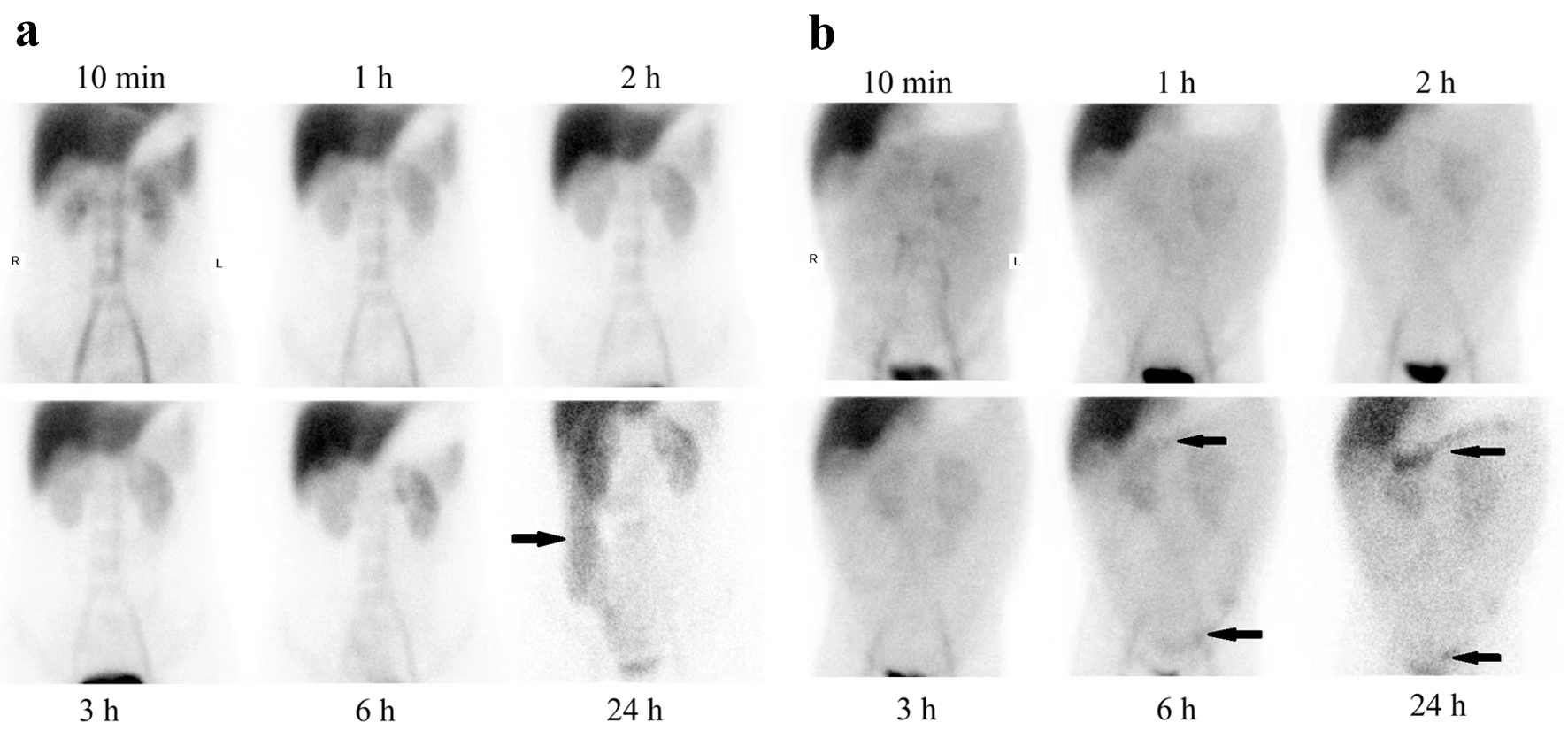
↓ Figure 1. 99mTc-HSA scintigraphy in two patients. Protein was lost from intestine (black arrowheads) in case 1 (a) and case 4 (b). HAS: human serum albumin.
| Gastroenterology Research, ISSN 1918-2805 print, 1918-2813 online, Open Access |
| Article copyright, the authors; Journal compilation copyright, Gastroenterol Res and Elmer Press Inc |
| Journal website https://gr.elmerpub.com |
Case Report
Volume 18, Number 3, June 2025, pages 152-158
Secondary Hyperparathyroidism in Primary Intestinal Lymphangiectasia: A Report of Four Cases
Figures
Table
| Case no. | Ca (mmol/L) | Albumin (g/L) | Adjusted Ca (mmol/L) | P (mmol/L) | Mg (mmol/L) | iPTH (pg/mL) | 25(OH)D (ng/mL) | ALP (U/L) | Urine Ca (mmol/24 h) | Urine P (mmol/24 h) |
|---|---|---|---|---|---|---|---|---|---|---|
| ALP: alkaline phosphatase; Ca: calcium; iPTH: intact parathyroid hormone; Mg: magnesium; 25(OH)D: 25-hydroxyvitamin D; P: phosphate. | ||||||||||
| 1 | 1.01 | 16.6 | 1.52 | 0.38 | 0.45 | 238.10 | < 3.0 | 1,107 | 6.34 | - |
| 2 | 1.03 | 16.2 | 1.506 | 0.79 | 0.47 | 230.4 | - | 311 | - | - |
| 3 | 1.36 | 16.1 | 1.838 | 1.14 | 0.52 | 202.50 | - | 103 | 3.11 | 1.43 |
| 4 | 1.98 | 34.8 | 2.08 | 1.48 | 0.83 | 302.9 | 3.0 | 238 | - | - |