Figures
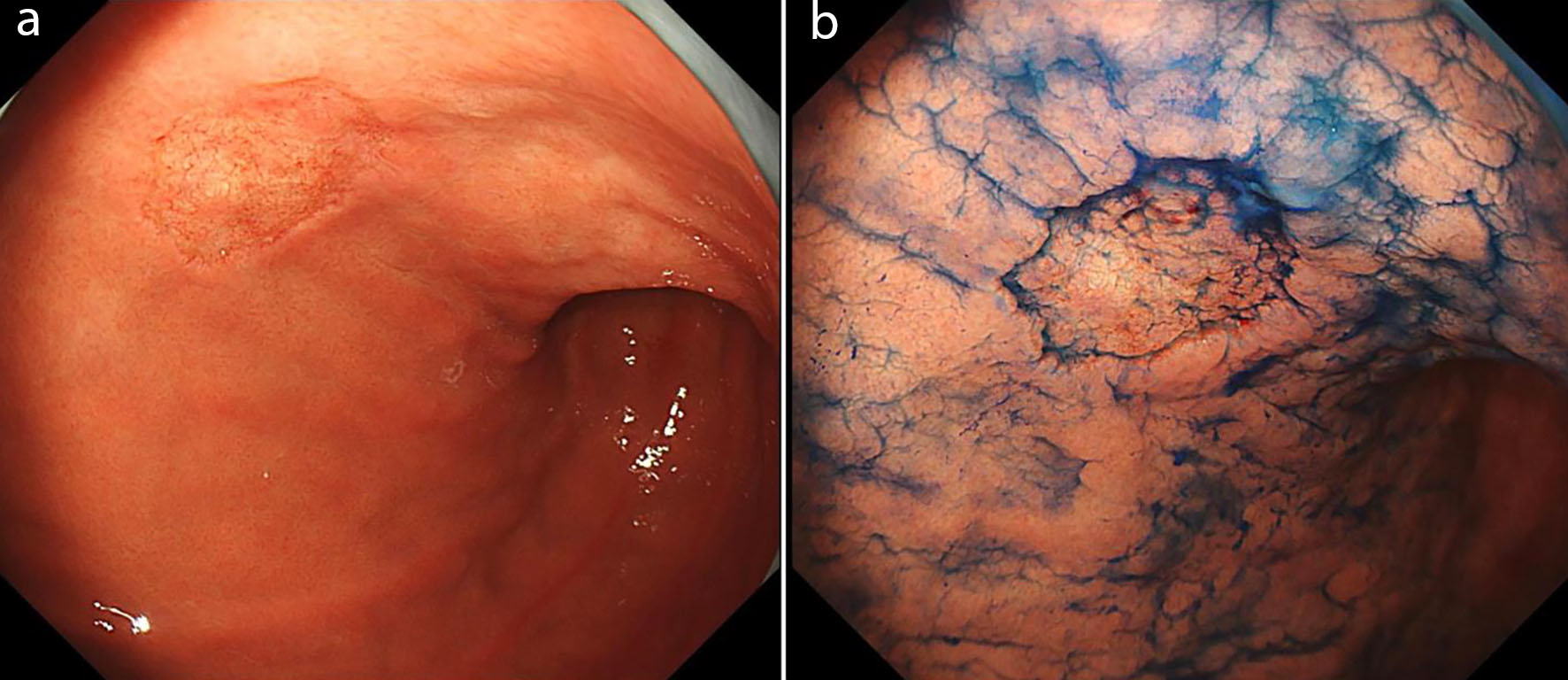
↓ Figure 1. A solitary 25-mm type 0-IIc lesion localized to the lesser curvature of the lower gastric body. (a) White-light endoscopy revealed an irregular, discolored depressed lesion. (b) Chromoendoscopy with indigo carmine demonstrated a coarse mucosal surface within the depressed area and an irregular “moth-eaten” appearance at the boundary between the lesion and the surrounding non-neoplastic mucosa.
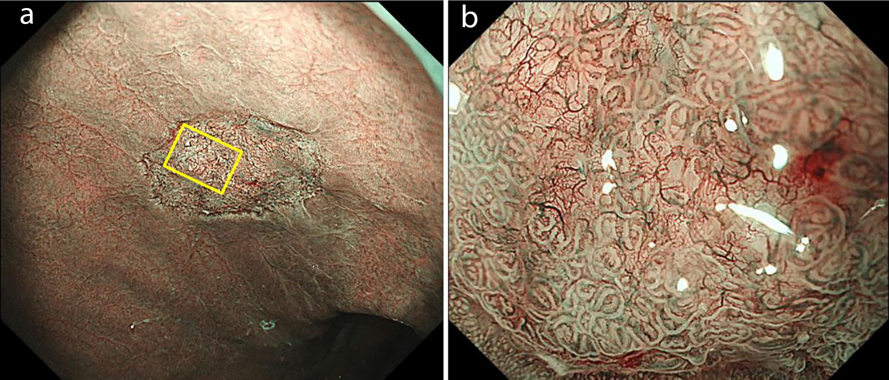
↓ Figure 2. Magnifying endoscopy with NBI. (a) A well-demarcated brownish area was observed. (b) Magnified view of the central part of the lesion showed obscured glandular structures with a mixture of normal-like architecture. Tortuous vessels were noted in areas with structural obscuration; however, no clearly atypical irregular microvascular patterns were identified. NBI: narrow-band imaging.
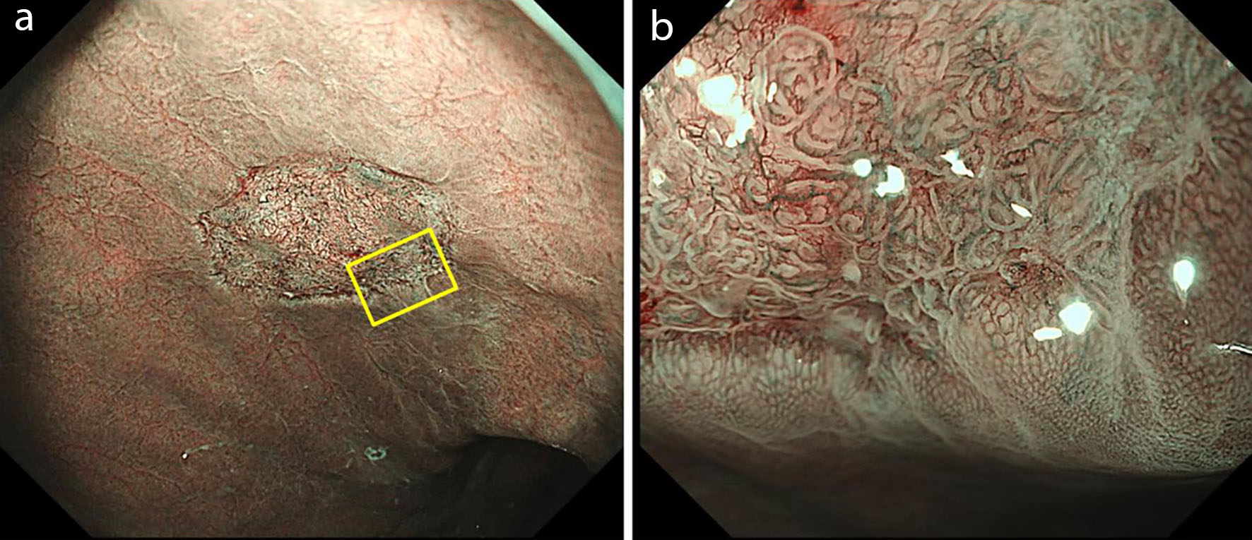
↓ Figure 3. Magnifying endoscopy with NBI of the lesion margin. (a) A clearly demarcated depressed change was observed at the margin of the lesion. (b) Within the lesion, enlarged glandular structures and prominent vascular patterns were observed compared to the surrounding mucosa; however, no definite irregular glandular or vascular architecture suggestive of malignancy was identified. NBI: narrow-band imaging.
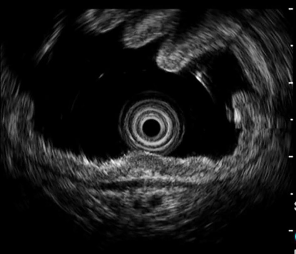
↓ Figure 4. Endoscopic ultrasonography using a 20-MHz probe demonstrated homogeneous hypoechoic thickening confined to the second layer, with no evidence of deeper submucosal invasion.
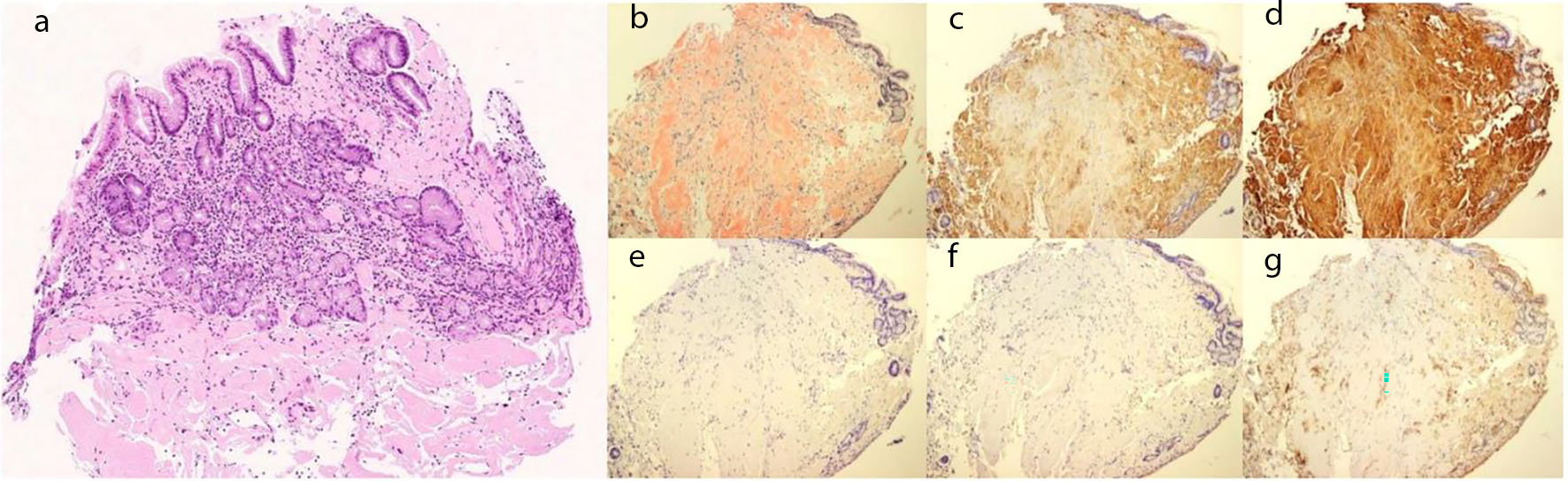
↓ Figure 5. Histopathological findings of biopsy specimens from the lesion. (a) No evidence of malignancy was observed, and amyloid deposition was identified mainly in the submucosa. (b) Congo red staining was positive for amyloid protein. (c) Immunohistochemical staining showed negativity for kappa light chain, (d) positivity for lambda light chain, (e) and negativity for amyloid A, (f) transthyretin, and (g) β2-microglobulin, confirming the diagnosis of AL (lambda-type) amyloidosis. AL: amyloid light chain.
Table
↓ Table 1. Reported Cases of Localized Gastric AL Amyloidosis Presenting With Depressed Lesions
| Author | Year | Age | Sex | Symptom | Location | Size (mm) | Endoscopic feature | NBI findings | Amyloid type | Underlying disease | Treatment |
|---|
| Mucosal structure | Microvasculature |
|---|
| AL: amyloid light chain; ND: not described; SMT: submucosal tumor; NBI: narrow-band imaging; RA: rheumatoid arthritis; EMR: endoscopic mucosal resection; ESD: endoscopic submucosal dissection; APC: argon plasma coagulation; AL-λ: amyloid light-chain lambda-type; AL-κ: amyloid light-chain kappa-type. |
| 1 | Yanai et al [6] | 1991 | 52 | F | None | Lower gastric body | 25 | Erosion | ND | ND | AL-λ | ND | EMR |
| 2 | Ebato et al [7] | 2012 | 77 | F | Anemia | Lower gastric body | 46 | Depressed | ND | ND | AL | ND | ESD |
| 3 | Yamaguchi et al [8] | 2015 | 49 | M | None | Lower gastric body | 15 | SMT + depressed | Groove-type surface pattern with an apparent demarcation line | ND | AL | ND | ND |
| 4 | Kagawa et al [9] | 2016 | 75 | M | None | Lower gastric body | 15 | Depressed | Irregular groove-type surface pattern | Round microvascular changes | AL | None | Follow-up |
| 5 | Ahn et al [10] | 2018 | 55 | F | None | Mid-gastric body | 20 | Depressed | ND | ND | AL-κ/λ | Breast cancer | Follow-up |
| 6 | Furukawa et al [11] | 2020 | 56 | M | None | Mid-gastric body | 40 | Depressed | Irregular groove-type surface pattern | Round small changes and dilated vessels without caliber | AL-κ | ND | Follow-up |
| 7 | Takahashi et al [12] | 2021 | 71 | F | None | Lower gastric body | 25 | SMT + depressed | No microsurface irregularity | Dilated vessels without microvessel irregularity | AL-λ | None | ESD |
| 8 | Hashimoto et al [13] | 2021 | 49 | M | None | Gastric antrum | 20 | SMT + depressed | ND | ND | AL-λ | None | Follow-up |
| 9 | Iwamuro et al [14] | 2023 | 58 | M | ND | Lower gastric body | 25 | Depressed | ND | ND | AL-λ | None | Follow-up |
| 10 | Iwamuro et al [14] | 2023 | 59 | M | ND | Lower gastric body | 20 | Depressed | Partial absence of microstructural patterns | Emphasized dilation of microvasculature | AL-λ | None | Follow-up |
| 11 | Iwamuro et al [14] | 2023 | 39 | M | ND | ND | 15 | Depressed | ND | ND | AL-κ | None | APC |
| 12 | Iwamuro et al [14] | 2023 | 50 | M | ND | Lower gastric body | 20 | Depressed | Partial absence of microstructural patterns | Emphasized dilation of microvasculature | AL-λ | None | Follow-up |
| 13 | Iwamuro et al [14] | 2023 | 51 | M | ND | ND | 35 | Depressed | ND | ND | AL-λ | None | Follow-up |
| 14 | Iwamuro et al [14] | 2023 | 62 | M | ND | Upper gastric body | 15 | Depressed | ND | ND | AL-κ | None | Follow-up |
| 15 | Present case | 2026 | 78 | F | None | Lower gastric body | 25 | Depressed | Partially preserved glandular structures | Tortuous vessels without definite irregularity | AL-λ | RA | Follow-up |