Figures
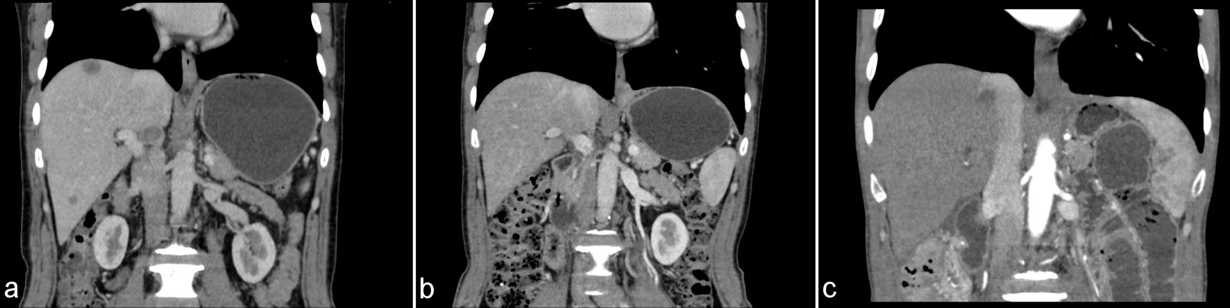
↓ Figure 1. Contrast-enhanced computed tomography (CT) images before and after systemic therapy. (a) Baseline (December 2025) CT showing gastric wall thickening and multiple hypodense liver lesions. (b) Restaging CT (March 2026) showing that the liver lesion is less distinct than before, suggesting that the lesion may be resolving. (c) Restaging CT (October 2026) showing no evidence of hepatic recurrence or new metastatic lesions.
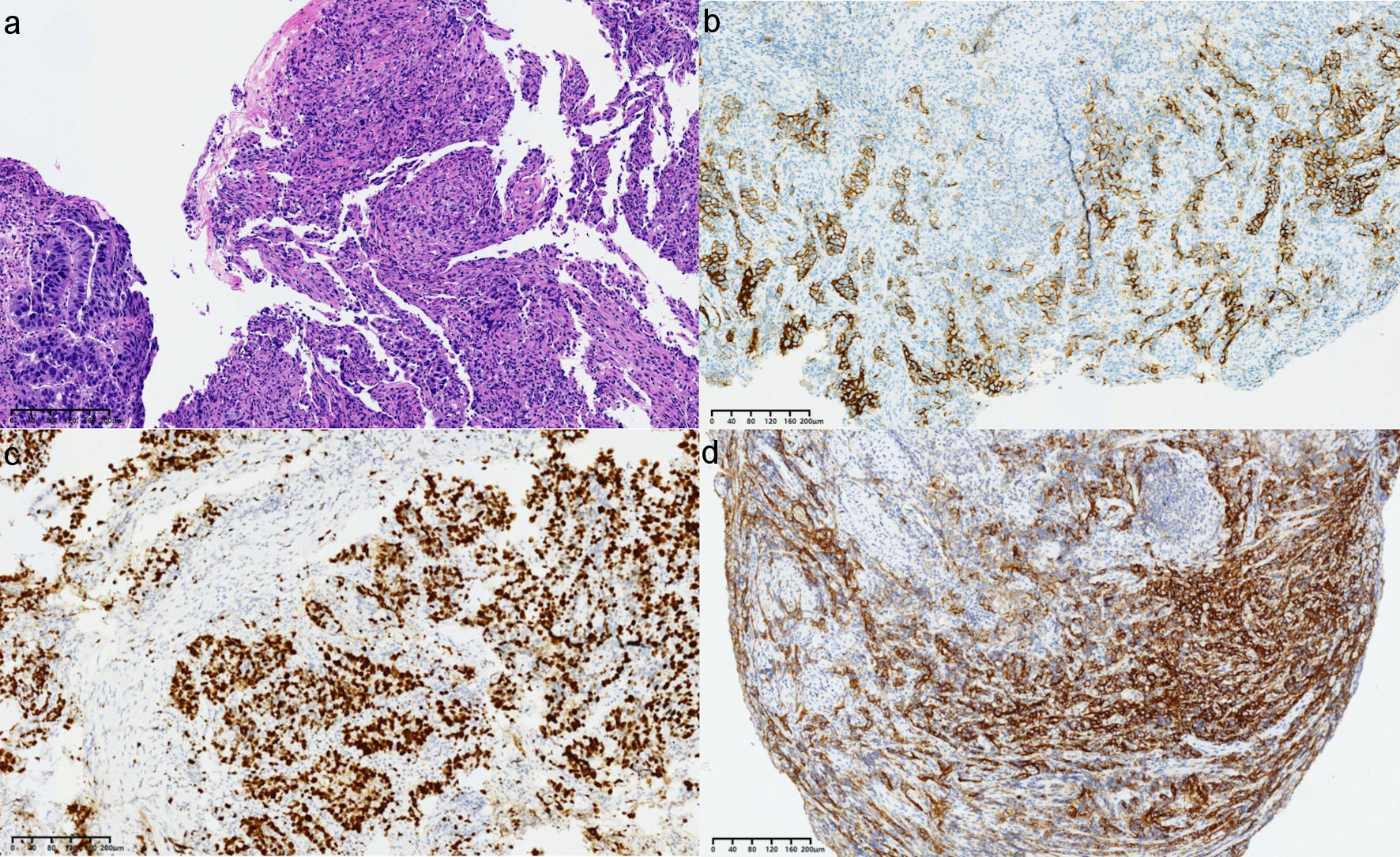
↓ Figure 2. Pre-treatment endoscopic biopsy of gastric carcinoma. (a) Hematoxylin and eosin (H&E) staining, × 10 magnification, poorly differentiated adenocarcinoma. (b) Human epidermal growth factor receptor 2 (HER2) immunohistochemistry (IHC), × 10, 3+ complete membranous staining. (c) Ki-67 proliferation index IHC, × 10, 90%. (d) Programmed death-ligand 1(22C3) IHC, × 10, combined positive score (CPS) = 30.
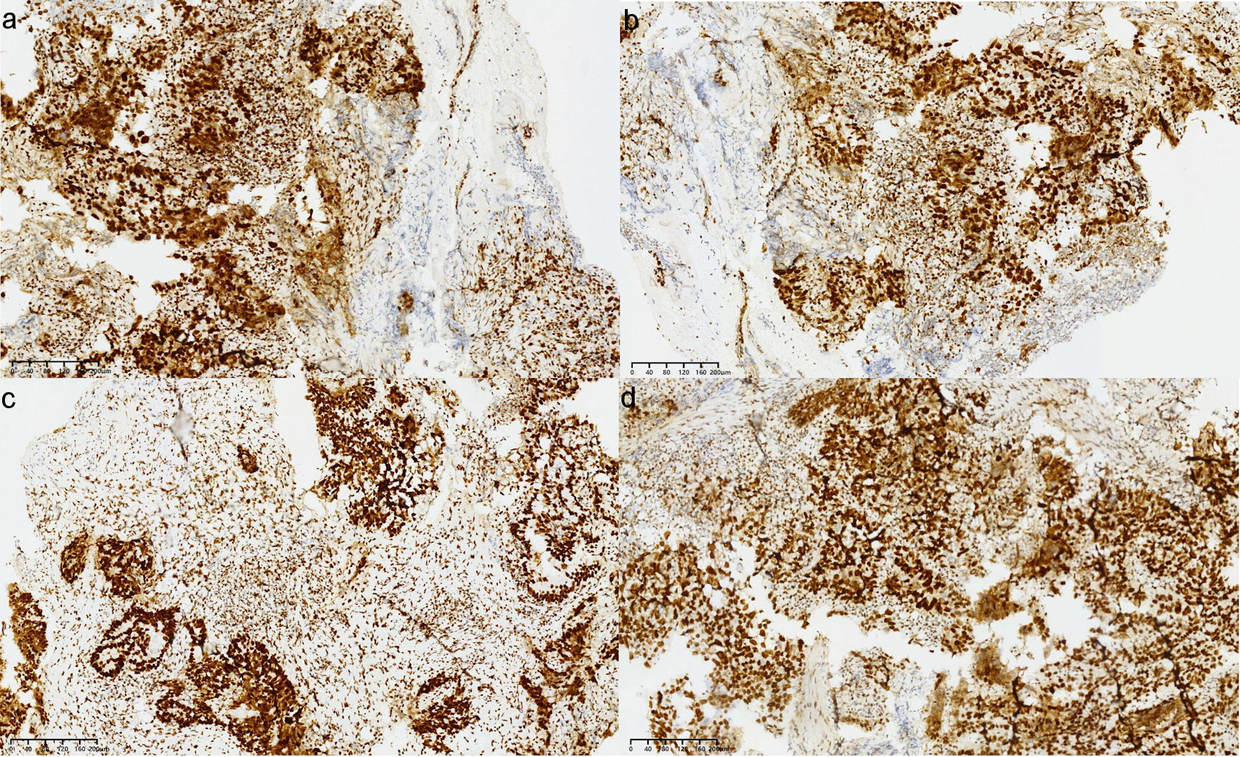
↓ Figure 3. Mismatch-repair protein immunohistochemistry of pre-treatment biopsy specimen (× 10). (a) mutL homolog 1 (MLH1) positive; (b) mutS homolog 2 (MSH2) positive; (c) mutS homolog 6 (MSH6) positive; (d) postmeiotic segregation increased 2 (PMS2) positive, consistent with proficient mismatch-repair (pMMR) status.
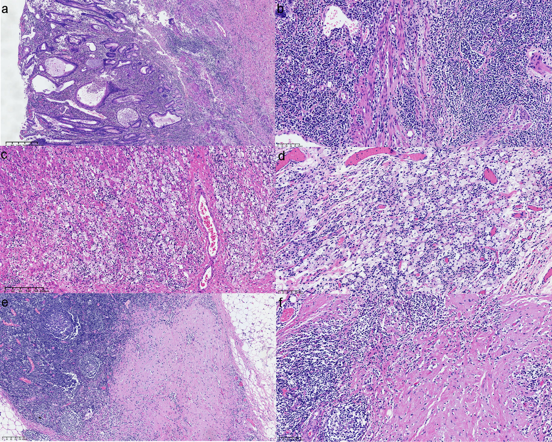
↓ Figure 4. Post-treatment surgical resection specimen showing complete pathological regression in the examined gastric and nodal tissue (tumor regression grade 0). (a, b) Primary tumor bed with extensive stromal fibrosis and inflammatory cell infiltration, no viable tumor cells identified (hematoxylin and eosin (H&E) staining; original magnification, × 10 (a) and × 20 (b)). (c, d) Foamy histiocytes and stromal fibrosis (H&E staining; original magnification, × 10 (c) and × 20 (d)). (e, f) Lymph node showing post-treatment reactive changes without metastatic carcinoma (H&E staining; original magnification, × 10 (e) and × 20 (f)).