| Gastroenterology Research, ISSN 1918-2805 print, 1918-2813 online, Open Access |
| Article copyright, the authors; Journal compilation copyright, Gastroenterol Res and Elmer Press Inc |
| Journal website https://gr.elmerpub.com |
Case Report
Volume 19, Number 5, October 2026, pages 288-294

Localized Gastric Amyloid Light Chain Amyloidosis With Atypical Depressed Morphology: Diagnostic Implications Based on Magnifying Endoscopy
Tadashi Miikea, Toshiki Kurokia, Yoshinori Ozonoa, Ryo Tsuzukib, Yoshiko Umekitab, Yuichiro Satob, Hiroshi Kawakamia, c
aDivision of Gastroenterology and Hepatology, Department of Internal Medicine, Faculty of Medicine, University of Miyazaki, Miyazaki, Japan
bSection of Oncopathology and Morphological Pathology, Department of Pathology, Faculty of Medicine, University of Miyazaki, Miyazaki, Japan
cCorresponding Author: Hiroshi Kawakami, Division of Gastroenterology and Hepatology, Department of Internal Medicine, Faculty of Medicine, University of Miyazaki, Miyazaki 889-1601, Japan
Manuscript submitted June 9, 2026, accepted August 26, 2026, published online October 31, 2026
Short title: Atypical Gastric AL Amyloidosis
doi: https://doi.org/10.14740/gr2176
| Abstract | ▴Top |
Gastrointestinal involvement of amyloidosis is uncommon and cases limited to the stomach with a depressed lesion are limited. Here, we report the case of a 78-year-old woman who had a solitary 25-mm depressed lesion with irregular and indistinct boundaries in the lower gastric body with esophagogastroduodenoscopy (EGD). Chromoendoscopy (CE) revealed a coarse mucosal surface with a “moth-eaten” appearance, suggesting early gastric cancer. Magnifying narrow-band imaging (NBI) revealed partially preserved glandular structures and tortuous vessels without definite irregular microvascular patterns (IMVPs). Endoscopic ultrasonography (EUS) revealed a homogeneous hypoechoic thickening confined to the superficial layers. Histopathological examination revealed amyloid deposits in the submucosa. Congo red staining was positive and immunohistochemical analysis confirmed amyloid light-chain lambda-type (AL-λ) deposition. Comprehensive systemic evaluation showed no evidence of systemic involvement, establishing a diagnosis of localized gastric immunoglobulin light chain amyloidosis. Further accumulation of similar cases is required to clarify the characteristic endoscopic findings and their clinical significance.
Keywords: Narrow-band imaging; Early gastric cancer; Lymphoma; Stomach
| Introduction | ▴Top |
Amyloidosis is a rare disorder of unknown etiology characterized by the extracellular deposition of amyloid fibrillar proteins in multiple organs, leading to organ dysfunction. It is categorized into systemic and localized forms, based on the distribution of amyloid deposits. Compared with systemic amyloidosis, cases of amyloid deposition confined to the gastrointestinal tract are relatively uncommon, and those limited to the stomach are rare. Furthermore, cases of depressed lesions are extremely rare.
In the gastrointestinal tract, amyloid proteins are most commonly of the amyloid light chain (AL) or amyloid A (AA) type and typically occur as part of a systemic disease with single-organ involvement [1]. Endoscopic features differ depending on the subtype. In AL, amyloid deposits accumulate from the muscularis mucosa to the muscularis propria in a nodular pattern, often presenting as thickened folds, multiple nodules, or submucosal tumor-like (SMT) elevations. In contrast, AA predominantly involves the lamina propria, resulting in a coarse mucosal surface [2].
In this case, amyloid deposition was localized in the stomach; however, the lesion exhibited a depressed morphology, which is not consistent with the typical endoscopic features of AL amyloidosis reported in the literature. Here, we report a rare case of localized gastric AL amyloidosis that presented as a depressed lesion requiring differentiation from early gastric cancer and lymphoma.
| Case Report | ▴Top |
A 78-year-old woman underwent esophagogastroduodenoscopy (EGD) at a local clinic in May 2024 as part of a routine health check-up. An irregular depressed lesion was detected in the lesser curvature of the lower gastric body. The patient was referred to our hospital for further evaluation and management.
The patient’s medical history included rheumatoid arthritis (RA), angina pectoris, hypertension, chronic obstructive pulmonary disease, and dyslipidemia. Her medications included irbesartan/amlodipine besylate, disopyramide phosphate, prasugrel hydrochloride, lansoprazole, brotizolam, atorvastatin calcium hydrate, and shakuyakukanzoto.
Repeat EGD at our institution revealed a solitary, localized 25-mm type 0-IIc lesion on the lesser curvature of the lower gastric body. On white-light endoscopy (WL), the lesion appeared as an irregular, discolored, and depressed area. Chromoendoscopy (CE) with indigo carmine demonstrated a coarse mucosal surface and an irregular boundary between the lesion and surrounding mucosa, showing a “moth-eaten” appearance suggesting depressed-type early gastric cancer (Fig. 1).
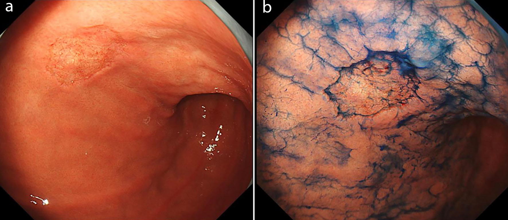 Click for large image | Figure 1. A solitary 25-mm type 0-IIc lesion localized to the lesser curvature of the lower gastric body. (a) White-light endoscopy revealed an irregular, discolored depressed lesion. (b) Chromoendoscopy with indigo carmine demonstrated a coarse mucosal surface within the depressed area and an irregular “moth-eaten” appearance at the boundary between the lesion and the surrounding non-neoplastic mucosa. |
Magnifying endoscopy with narrow-band imaging (NBI) revealed partially obscured glandular structures with a combination of an enlarged, normal-like architecture. Tortuous vessels were observed in areas with unclear structures; however, no definite atypical or irregular microvascular patterns (IMVPs) suggestive of malignancy were observed (Figs. 2 and 3).
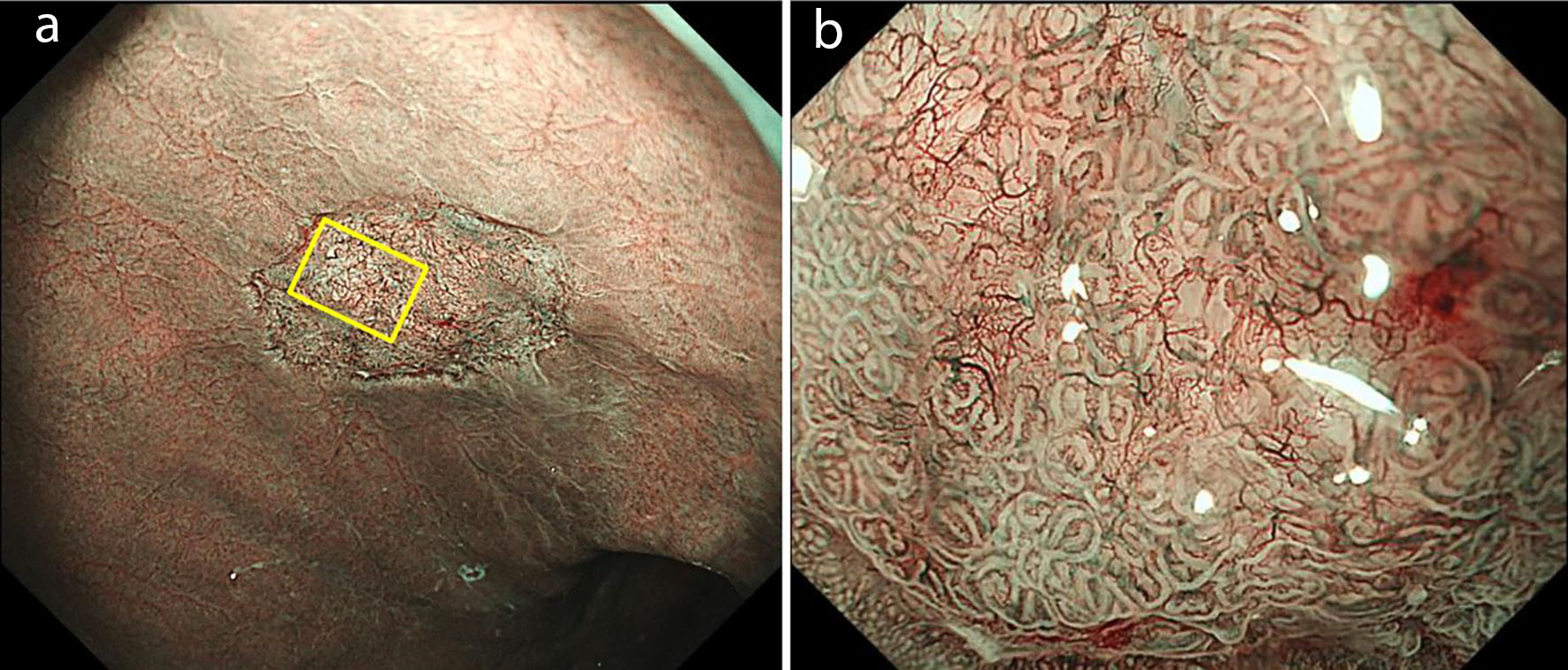 Click for large image | Figure 2. Magnifying endoscopy with NBI. (a) A well-demarcated brownish area was observed. (b) Magnified view of the central part of the lesion showed obscured glandular structures with a mixture of normal-like architecture. Tortuous vessels were noted in areas with structural obscuration; however, no clearly atypical irregular microvascular patterns were identified. NBI: narrow-band imaging. |
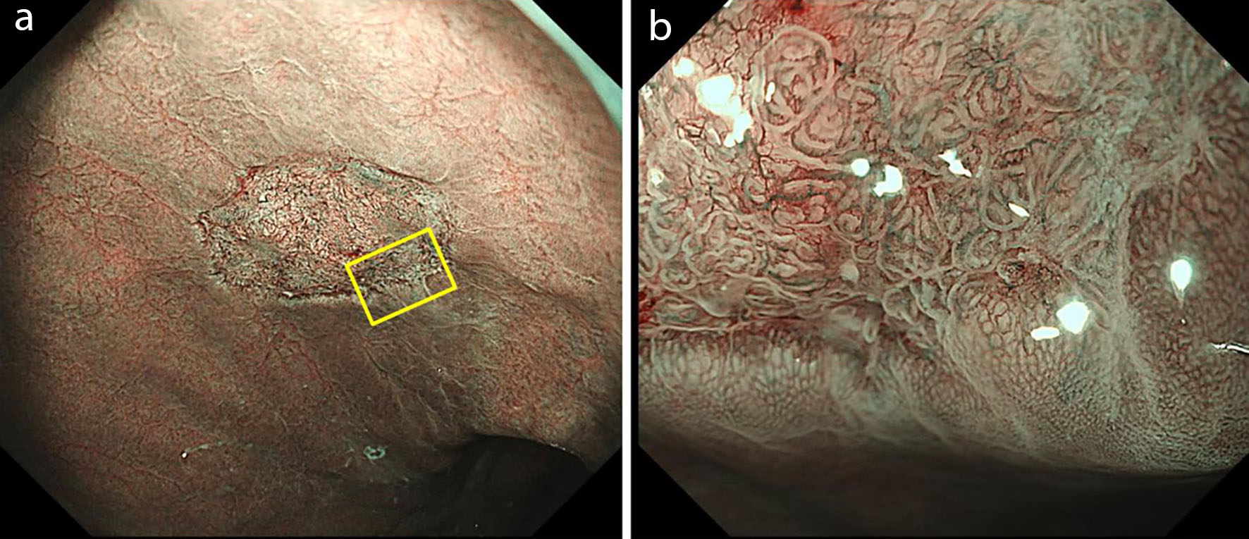 Click for large image | Figure 3. Magnifying endoscopy with NBI of the lesion margin. (a) A clearly demarcated depressed change was observed at the margin of the lesion. (b) Within the lesion, enlarged glandular structures and prominent vascular patterns were observed compared to the surrounding mucosa; however, no definite irregular glandular or vascular architecture suggestive of malignancy was identified. NBI: narrow-band imaging. |
Considering the possibility of early gastric cancer or mucosa-associated lymphoid tissue (MALT) lymphoma, endoscopic ultrasonography (EUS) was performed using a 20-MHz probe. This demonstrated a homogeneous hypoechoic thickening confined to the second layer (Fig. 4), suggesting no evidence of deep submucosal invasion.
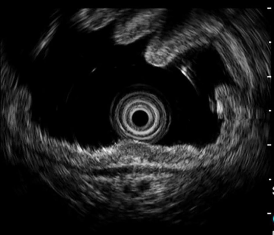 Click for large image | Figure 4. Endoscopic ultrasonography using a 20-MHz probe demonstrated homogeneous hypoechoic thickening confined to the second layer, with no evidence of deeper submucosal invasion. |
Histopathological examination of biopsy specimens obtained from the lesion margin showed no evidence of malignancy but revealed amyloid deposition in the submucosa. Congo red staining was positive, and immunohistochemical analysis confirmed amyloid light-chain lambda-type (AL-λ) deposition (Fig. 5).
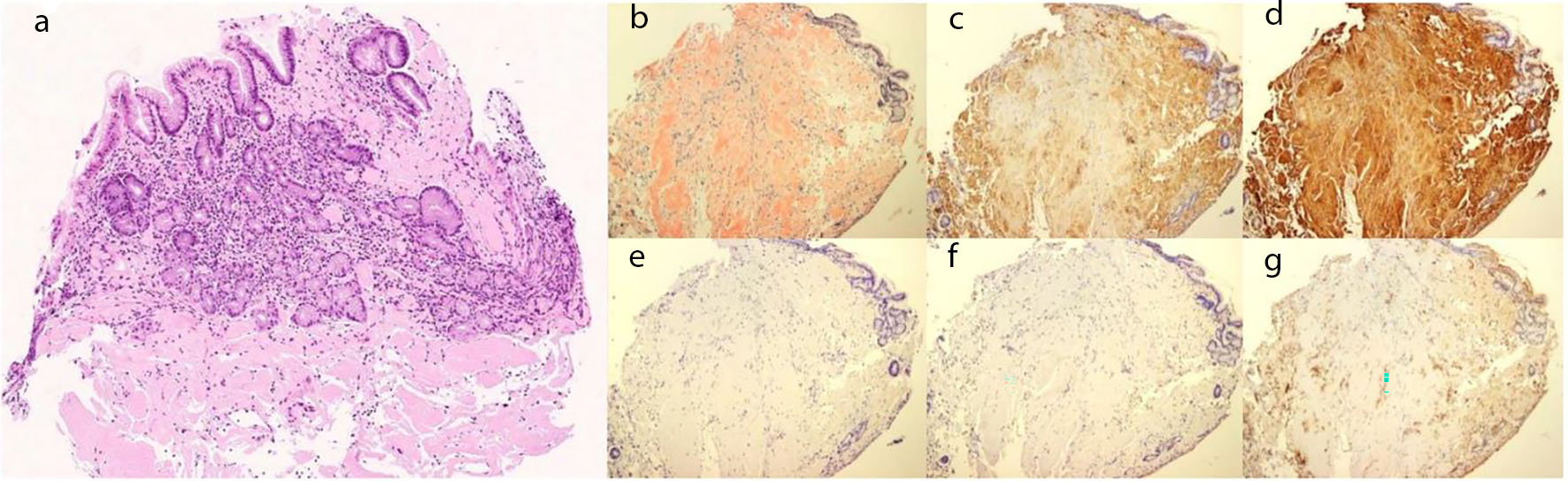 Click for large image | Figure 5. Histopathological findings of biopsy specimens from the lesion. (a) No evidence of malignancy was observed, and amyloid deposition was identified mainly in the submucosa. (b) Congo red staining was positive for amyloid protein. (c) Immunohistochemical staining showed negativity for kappa light chain, (d) positivity for lambda light chain, (e) and negativity for amyloid A, (f) transthyretin, and (g) β2-microglobulin, confirming the diagnosis of AL (lambda-type) amyloidosis. AL: amyloid light chain. |
To exclude systemic amyloidosis, a comprehensive evaluation was performed in collaboration with the Departments of Cardiology, Hematology, and Radiology. These included chest and abdominal computed tomography, abdominal ultrasonography, electrocardiography, echocardiography, serum protein electrophoresis, urine and serum immunoelectrophoresis, bone marrow aspiration, G-band analysis, and colonoscopy. No findings suggestive of systemic amyloidosis were observed. No amyloid deposition was observed in the colon and duodenum.
Therefore, localized gastric AL amyloidosis was diagnosed. Furthermore, AA amyloidosis associated with RA was considered unlikely, based on the patient’s medical history. Currently, there is no evidence of cardiac dysfunction or multiple myeloma. The patient and her family were informed of the diagnosis, and careful follow-up was continued at our hospital and the referring clinic. Subsequent examinations revealed no changes in the lesions.
| Discussion | ▴Top |
Primary amyloidosis is a metabolic disorder of unknown etiology, characterized by the extracellular deposition of amyloid fibrils in various organs, resulting in varying clinical manifestations. The gross endoscopic findings of gastrointestinal amyloidosis are variable and include fine granular mucosa, elevated lesions, ulcerations, mucosal fragility, and wall thickening [3, 4]. Menke et al reported that only eight of 769 cases (1%) of gastrointestinal amyloidosis were diagnosed by biopsy, suggesting that diagnosis is challenging unless systemic amyloidosis has already been identified [5].
In this case, although the lesion was localized in the stomach, it exhibited an atypical depressed morphology that differed from the prototypical elevated or nodular appearance generally reported in AL amyloidosis. Importantly, the lesion closely mimicked early gastric cancer and lymphoma, highlighting the diagnostic difficulty during routine endoscopy. Similar cases are extremely rare; a PubMed search identified 14 previously reported cases, and our case was added to these cases for a total of 15 cases (Table 1) [6–14]. The median age of the patients was 56 years (range, 39–78 years), most were men (66.7%), and the lower gastric body was the most frequently involved site (60%). Among cases in which the light-chain subtype was reported, AL-λ was predominant (66.7%). Among the 15 cases, including our case, nine patients (60.0%) were asymptomatic, whereas one patient (6.7%) presented with anemia.
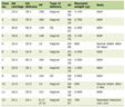 Click to view | Table 1. Reported Cases of Localized Gastric AL Amyloidosis Presenting With Depressed Lesions |
According to Iwamuro et al [14], localized gastric AL amyloidosis can present with various morphological features, including SMT elevations, erythema, and depressed lesions, making diagnosis based on morphology alone challenging. An irregular depressed lesion, such as that observed in this case, closely resembles early gastric cancer or lymphoma, necessitating careful differential diagnosis.
Image-enhanced endoscopy (IEE), including magnifying NBI, may be useful for differentiating these lesions. Early gastric cancer typically demonstrates a distinct demarcation line and IMVPs characterized by dilation, tortuosity, heterogeneity, and asymmetrical distribution [15]. In contrast, lymphomas, such as MALT lymphoma, are characterized by a tree-like appearance (TLA) of abnormal branching vessels in areas of glandular destruction [16]. A review of previously reported cases revealed that magnifying NBI findings in localized gastric amyloidosis are variable and remain poorly characterized. Reported mucosal surface findings include groove-type or irregular groove-type surface patterns, partial absence of microstructural patterns, and preserved micro-surface structures, whereas vascular findings include round microvascular changes and dilated vessels without marked caliber irregularities. However, destructive or highly atypical vascular patterns suggestive of malignancy have not been observed.
In the present case, magnifying NBI demonstrated partially preserved glandular structures with tortuous vessels, with no definite IMVPs or tree-like abnormal vessels. Although the lesion showed some structural alterations, the overall glandular architecture was relatively maintained, and vascular atypia was minimal compared with that typically observed in gastric cancer or lymphoma. Collectively, these findings suggest that magnifying NBI may provide clinically useful clues for differentiating localized gastric amyloidosis from malignant depressed lesions before histological confirmation. However, histological confirmation by biopsy and immunohistochemical evaluation remain essential for a definitive diagnosis.
Regarding the mechanism of depressed morphology, amyloid deposition within the vascular walls leads to luminal narrowing, vascular fragility, and impaired blood flow. This may result in ischemic changes, tissue atrophy, and decreased gastric wall elasticity, leading to the formation of irregular depressed lesion [17]. Localized gastric AL amyloidosis generally has a favorable clinical course and is often managed conservatively [14]. Among the 15 reported cases, including our case, 10 patients (66.7%) were managed with follow-up alone, whereas four patients (26.7%) underwent endoscopic treatment, including endoscopic submucosal dissection (ESD), endoscopic mucosal resection (EMR), or argon plasma coagulation (APC, Table 1). Treatment information was not described in one case. Careful observation is appropriate in asymptomatic patients with localized disease, as in this case. However, treatment may be indicated in cases with complications, such as bleeding, lesion enlargement, or suspicion of malignancy. Although the patient has been followed for approximately 2 years and remains asymptomatic, careful long-term follow-up is warranted because morphological changes have been reported during long-term observation [12].
This study had several limitations. This is a single-case report, and the mechanism underlying the localized deposition and atypical depressed morphology remains unclear. Considering the limited number of reported cases, further accumulation and analysis of similar cases are necessary to better understand the clinicopathological features and prognosis of this condition.
Learning points
Overall, we report a rare case of localized gastric AL amyloidosis presenting as an atypical depressed lesion closely mimicking early gastric cancer and lymphoma. Although amyloidosis is generally a systemic disease, localized involvement of the stomach is uncommon, and depressed-type lesions are rare.
This case highlights the diagnostic challenges posed by atypical endoscopic findings. Morphological assessment using conventional endoscopy alone may be insufficient to distinguish localized gastric amyloidosis from malignant lesions. Magnifying endoscopy with NBI can provide important diagnostic clues, as the absence of clearly detected IMVPs and presence of partially preserved glandular structures may suggest a non-neoplastic process.
Histopathological evaluation is essential for a definitive diagnosis in cases with suspicious or ambiguous endoscopic features. A comprehensive systemic workup is required to exclude systemic amyloidosis although the lesion appears to be localized.
Although localized gastric AL amyloidosis is generally associated with a favorable prognosis, careful long-term follow-up is warranted. Clinicians should be aware of the potential risk of gastrointestinal bleeding due to vascular fragility caused by amyloid deposition.
Acknowledgments
We express our sincere gratitude to Dr. TA from the Department of Pathology, Tokyo Metropolitan Institute for Geriatrics and Gerontology, Tokyo, Japan, for his valuable assistance with special staining and pathological evaluation.
Financial Disclosure
None to declare.
Conflict of Interest
None to declare.
Informed Consent
Informed consent was obtained for the use of deidentified information for publication. This case report followed the guidelines of the Institutional Review Board of University of Miyazaki Hospital, Miyazaki, Japan.
Author Contributions
Concept, drafting, and final review, TM; direct patient care, TK; pathological evaluation, RT, YU, and YS; manuscript revision, YO; critical review and supervision, HK. All authors approved the final manuscript.
Data Availability
The data supporting the findings of this study are available from the corresponding author upon reasonable request.
Abbreviations
AA: amyloid A; AL: amyloid light chain; CE: chromoendoscopy; EGD: esophagogastroduodenoscopy; EMR: endoscopic mucosal resection; ESD: endoscopic submucosal dissection; EUS: endoscopic ultrasonography; IMVP: irregular microvascular pattern; MALT: mucosa-associated lymphoid tissue; NBI: narrow-band imaging; RA: rheumatoid arthritis; SMT: submucosal tumor; TLA: tree-like appearance; WL: white-light endoscopy
| References | ▴Top |
- Gilat T, Revach M, Sohar E. Deposition of amyloid in the gastrointestinal tract. Gut. 1969;10(2):98-104.
doi pubmed - Hokama A, Kishimoto K, Nakamoto M, Kobashigawa C, Hirata T, Kinjo N, Kinjo F, et al. Endoscopic and histopathological features of gastrointestinal amyloidosis. World J Gastrointest Endosc. 2011;3(8):157-161.
doi pubmed - Bighi S, Trevisani L, Lupi L, Ariutti R, Cervi PM, Liboni A. Amyloidosis of the gastric stump: radiographic and CT findings. Gastrointest Radiol. 1990;15(3):197-198.
doi pubmed - Ueda M, Horibata Y, Shono M, Misumi Y, Oshima T, Su Y, Tasaki M, et al. Clinicopathological features of senile systemic amyloidosis: an ante- and post-mortem study. Mod Pathol. 2011;24(12):1533-1544.
doi pubmed - Menke DM, Kyle RA, Fleming CR, Wolfe JT, 3rd, Kurtin PJ, Oldenburg WA. Symptomatic gastric amyloidosis in patients with primary systemic amyloidosis. Mayo Clin Proc. 1993;68(8):763-767.
doi pubmed - Yanai H, Fuji T, Tada M, Aibe T, Karita M, Fujimura H, Okita K, et al. Localized amyloidosis of the stomach. Gastrointest Endosc. 1991;37(3):408-409.
doi pubmed - Ebato T, Yamamoto T, Abe K, Ishii T, Kuyama Y. Endoscopic removal of localized gastric amyloidosis. Endoscopy. 2012;44(Suppl 2 UCTN):E351.
doi pubmed - Yamaguchi T, Inoue T, Nishida T, Kato M, Hayashi Y, Tsujii Y, Maekawa A, et al. Localized gastric amyloidosis mimicking a submucosal tumor-like gastric cancer. Gastrointest Endosc. 2015;82(1):175-177.
doi pubmed - Kagawa M, Fujino Y, Muguruma N, Murayama N, Okamoto K, Kitamura S, Kimura T, et al. Localized amyloidosis of the stomach mimicking a superficial gastric cancer. Clin J Gastroenterol. 2016;9(3):109-113.
doi pubmed - Ahn YH, Rhee YY, Choi SC, Seo GS. Localized gastric amyloidosis with Kappa and Lambda Light Chain Co-Expression. Clin Endosc. 2018;51(3):285-288.
doi pubmed - Furukawa K, Miyahara R, Funasaka K, Fujishiro M. Localized gastric amyloidosis. J Gastrointestin Liver Dis. 2020;29(4):497.
doi pubmed - Takahashi Y, Hatta W, Koike T, Ogata Y, Fujiya T, Saito M, Jin X, et al. Localized gastric amyloidosis that displayed morphological changes over 10 years of observation. Intern Med. 2021;60(4):539-543.
doi pubmed - Hashimoto C, Hikichi T, Hashimoto M, Waragai Y, Takasumi M, Nakamura J, Kato T, et al. Localized gastric amyloidosis diagnosed by endoscopic ultrasound-guided fine-needle aspiration. Clin J Gastroenterol. 2021;14(4):1036-1041.
doi pubmed - Iwamuro M, Tanaka S, Toyokawa T, Nishimura M, Tsuzuki T, Miyahara K, Negishi S, et al. Endoscopic manifestations and clinical characteristics of localized gastric light-chain amyloidosis. Acta Med Okayama. 2023;77(5):545-552.
doi pubmed - Yamada S, Doyama H, Yao K, Uedo N, Ezoe Y, Oda I, Kaneko K, et al. An efficient diagnostic strategy for small, depressed early gastric cancer with magnifying narrow-band imaging: a post-hoc analysis of a prospective randomized controlled trial. Gastrointest Endosc. 2014;79(1):55-63.
doi pubmed - Nonaka K, Ishikawa K, Arai S, Nakao M, Shimizu M, Sakurai T, Nagata K, et al. A case of gastric mucosa-associated lymphoid tissue lymphoma in which magnified endoscopy with narrow band imaging was useful in the diagnosis. World J Gastrointest Endosc. 2012;4(4):151-156.
doi pubmed - Hirschfield GM. Amyloidosis: a clinico-pathophysiological synopsis. Semin Cell Dev Biol. 2004;15(1):39-44.
doi pubmed
This article is distributed under the terms of the Creative Commons Attribution 4.0 International License (CC BY 4.0), which permits unrestricted use, distribution, and reproduction in any medium, including commercial use, provided the original work is properly cited.
Gastroenterology Research is published by Elmer Press Inc.