| Gastroenterology Research, ISSN 1918-2805 print, 1918-2813 online, Open Access |
| Article copyright, the authors; Journal compilation copyright, Gastroenterol Res and Elmer Press Inc |
| Journal website https://gr.elmerpub.com |
Original Article
Volume 000, Number 000, November 2026, pages 000-000

Non-Surgical Esophageal Injury in Critically Ill Patients: Clinical Characteristics, Complications, and Interdisciplinary Management
Philipp Kazuo Omuroa, d, Dominique Harta, Seung-Hun Chonb, Dolores T. Kraussb, Hans F. Fuchsb, Wolfgang A. Wetscha, Christoph Neumann-Haefelinc, Christiane J. Brunsb, Andrea U. Steinbickera, Fabian Dussea
aDepartment of Anesthesiology and Intensive Care Medicine, Faculty of Medicine and University Hospital of Cologne, University of Cologne, Cologne, Germany
bDepartment of General, Visceral, Thoracic and Transplantation Surgery, Faculty of Medicine and University Hospital of Cologne, University of Cologne, Cologne, Germany
cDepartment of Gastroenterology and Hepatology, Faculty of Medicine and University Hospital of Cologne, University of Cologne, Cologne, Germany
dCorresponding Author: Philipp Kazuo Omuro, Department of Anesthesiology and Intensive Care Medicine, Faculty of Medicine and University Hospital of Cologne, University of Cologne, 50937 Cologne, Germany
Manuscript submitted July 14, 2026, accepted September 10, 2026, published online November 10, 2026
Short title: Esophageal Injury in Critically Ill Patients
doi: https://doi.org/10.14740/gr2180
| Abstract | ▴Top |
Background: Non-surgical esophageal injury can cause mediastinal or pleural infection, organ dysfunction, and prolonged critical care. Evidence in critically ill patients regarding perforation status, diagnostic pathways, and treatment remains limited. We described clinical characteristics, complications, management, and outcomes and compared patients with and without confirmed perforation.
Methods: We retrospectively analyzed 42 adults with non-surgical esophageal injury treated for more than 24 h in a surgical intensive care unit between 2017 and 2023. Patients were compared by confirmed perforation. Categorical variables were analyzed using chi-square or Fisher’s exact tests, continuous variables using the Mann–Whitney U test, and exploratory associations using Spearman’s rank correlation.
Results: Mechanical trauma was most common (83.3%); 22 patients (52.4%) had perforation. Endoscopy was performed in 97.6% and computed tomography in 78.6%. Compared with patients without perforation, those with perforation had more sepsis (45.5% vs. 0.0%), septic shock (31.8% vs. 0.0%), acute kidney injury (27.3% vs. 0.0%), mediastinitis (50.0% vs. 5.0%), and pleural empyema (36.4% vs. 0.0%). Intensive care unit stay was longer with perforation (12.0 (5.8–22.3) vs. 4.5 (2.8–8.0) days; P = 0.005). Intracavitary endoscopic vacuum therapy was used in 19 patients, including two who also received a stent; surgery in 15; and both treatments in eight. Sixteen received supportive care without defect-directed intervention. Endoscopic vacuum therapy started within 12 h of intensive care unit admission in 18/19 patients and within 16 h in all. In-hospital mortality was 2.4% (n = 1).
Conclusions: Non-surgical esophageal injury is heterogeneous. Confirmed perforation was associated with greater morbidity and longer intensive care treatment. Early computed tomography and endoscopy, repeated interdisciplinary reassessment, and individualized supportive, endoscopic, or surgical therapy are central to management.
Keywords: Esophageal injury; Esophageal perforation; Critical care; Mediastinitis; Endoscopy; Interdisciplinary management
| Introduction | ▴Top |
Non-surgical injury of the esophagus is rare but potentially life-threatening because the esophagus traverses the neck, thorax, and abdomen in close proximity to vital structures. Etiologies include iatrogenic injury, foreign-body trauma, external trauma, caustic ingestion, spontaneous perforation, and other forms of mechanical injury. Clinical presentation ranges from limited mucosal injury to full-thickness perforation with mediastinal or pleural contamination, organ dysfunction, and septic shock [1–3].
The diagnostic objectives are to identify the lesion, determine whether transmural perforation is present, define the extent of contamination, and assess adjacent structures. Computed tomography (CT) and endoscopy provide complementary anatomical and mucosal information, while treatment may comprise supportive care, endoscopic therapy, image-guided drainage, surgery, or a combination of these approaches [4–8]. Contemporary evidence emphasizes individualized multidisciplinary management and treatment in centers with appropriate endoscopic, surgical, radiological, and critical care expertise [3, 8].
Because of the rarity and heterogeneity of non-surgical esophageal injury, current evidence is derived mainly from mixed cohorts of perforation or trauma. Data focusing specifically on critical care requirements, organ dysfunction, airway management, infection control, treatment timing, and overlapping interdisciplinary treatment pathways remain limited [3, 6, 8].
This study aimed to describe the clinical characteristics, diagnostic pathways, critical care requirements, therapeutic strategies, and outcomes of critically ill patients with non-surgical esophageal injury, with particular focus on differences between patients with and without confirmed esophageal perforation.
| Materials and Methods | ▴Top |
Study design and population
We performed a retrospective single-center cohort study of adult patients treated at the University Hospital of Cologne between 2017 and 2023. Cases were identified through an ICD-10-code-based database search in eisTIK using prespecified codes for esophageal injury (Supplementary Material 1, gr.elmerpub.com). Patients were eligible if they were 18 years or older, had a non-surgical esophageal injury, and received treatment in a surgical intensive care unit (ICU) for more than 24 h. Patients younger than 18 years, patients without ICU treatment, and patients with surgery-related or operative esophageal injuries were excluded.
Confirmed esophageal perforation was defined as a full-thickness disruption of the esophageal wall demonstrated by at least one of the following: direct visualization of a transmural defect during endoscopy or surgery, extraluminal leakage of orally administered contrast on CT, or a clearly demonstrated communication between the esophageal lumen and an adjacent mediastinal or pleural collection. Indirect findings alone, such as pneumomediastinum, pleural effusion, or mediastinal inflammation without a demonstrable defect or leak, were not considered sufficient. Classification was based on the final interdisciplinary assessment documented in the medical record.
Treatment definitions, selection, and timing
Treatment decisions were individualized in interdisciplinary consultation without a prespecified algorithm. Factors considered included the presence and extent of perforation, defect size and location, endoscopic accessibility, tissue viability, mediastinal or pleural contamination, mediastinitis or pleural empyema, clinical instability, sepsis, response to initial treatment, and failure of a preceding strategy. Intracavitary endoscopic vacuum therapy (EVT) was the preferred primary endoscopic defect treatment when technically feasible. Esophageal stents were used sparingly in individually selected cases. Surgery was considered particularly when adequate source control was unlikely to be achieved endoscopically, when contamination or tissue damage was extensive, in clinically unstable or septic patients, or after failure of less invasive therapy. Supportive-only management was selected when interdisciplinary assessment identified no indication for defect-directed EVT or surgery.
All patients received supportive and adjunctive care irrespective of whether EVT or surgery was added. Depending on individual requirements, this comprised nil per os, anti-infective therapy, proton pump inhibition, enteral feeding, and decompression using a triple-lumen tube, parenteral nutrition, and pleural or mediastinal drainage. Because modalities could be combined or applied sequentially, EVT, esophageal stenting, surgery, and supportive care were not treated as inherently exclusive categories. For the mutually exclusive treatment-pattern analysis, supportive care without defect-directed intervention was defined as supportive management without EVT or surgery.
The exact onset of injury or perforation was frequently uncertain, particularly in patients transferred from other hospitals. Treatment times were therefore calculated from ICU admission at our center rather than from the presumed time of injury. The first defect-directed intervention was defined as the earlier of EVT initiation or surgery, and modality-specific timing summaries included only patients who received the respective treatment. Outcomes were assessed during the index hospitalization. Systematic post-discharge follow-up was not available.
Institutional Review Board Approval and Ethical Compliance
The study was reviewed by the Ethics Committee of the Medical Faculty of the University of Cologne, which determined that formal ethical approval was not required because of the retrospective design and the use of fully anonymized patient data (Reference Number 24-1075-retro). The requirement for informed consent was waived. According to Section 15 of the Professional Code for Physicians in North Rhine and Recital 26 of the European Union General Data Protection Regulation, ethical approval and informed consent are not required for research involving anonymous data when no personal identifiers are collected or processed and re-identification is not possible. The study was conducted in compliance with applicable institutional ethical standards and the Declaration of Helsinki.
Statistical analysis
Statistical analyses were performed using IBM SPSS Statistics 29 (IBM Corp., Armonk, NY, USA). Continuous variables are reported as median (interquartile range (IQR)) and were compared using the Mann–Whitney U test. Categorical variables are reported as n (%) and were compared using the chi-square test or Fisher’s exact test, as appropriate. Exploratory associations were assessed using Spearman’s rank correlation. All tests were two-sided, and P < 0.05 was considered statistically significant. Given the exploratory nature and small sample size, no adjustment for multiple testing was performed.
| Results | ▴Top |
Study population
A total of 267 datasets were screened. No patients were younger than 18 years; 65 patients did not require ICU treatment for at least 24 h, 56 had surgery-related esophageal lesions, and 104 duplicate datasets were excluded. After all exclusion criteria were applied, 42 patients were included in the final analysis.
Patient characteristics and injury patterns
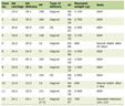
Patient characteristics, injury patterns, and direct complications are shown in Tables 1 and 2.
 Click to view | Table 1. Patient Characteristics and Comorbidities |
Click to view | Table 2. Characteristics of Esophageal Injury and Associated Complications |
Critical care course and organ dysfunction
The median ICU length of stay was 8 (3–19) days, and the median hospital length of stay was 21 (11–33) days. ICU length of stay was longer in patients with perforation (12.0 (5.8–22.3) vs. 4.5 (2.8–8.0) days; U = 330.5; P = 0.005), whereas hospital length of stay showed no clear between-group difference (25.0 (13.0–50.0) vs. 17.0 (10.3–22.8) days; U = 251.0; P = 0.083). The association between perforation and ICU length of stay was also supported by exploratory Spearman rank correlation (rho = 0.436; P = 0.004).
Sepsis occurred in 10 patients (23.8%), septic shock in seven (16.7%), and acute kidney injury in six (14.3%). All of these events occurred in patients with confirmed perforation: sepsis, 45.5% vs. 0.0% (P < 0.001); septic shock, 31.8% vs. 0.0% (P = 0.009); and acute kidney injury, 27.3% vs. 0.0% (P = 0.022). Invasive ventilation was more frequent in patients with perforation (77.3% vs. 40.0%; P = 0.027). Vasopressors were required in 59.5% of patients and inotropic agents in 7.1%. Respiratory support and pulmonary complications are summarized in Tables 3 and 4.
Click to view | Table 3. Respiratory Support and Airway Interventions During the Intensive Care Unit Stay |
Click to view | Table 4. Pulmonary and Respiratory Complications |
At ICU admission, median C-reactive protein was 82.6 (10.3–192.9) mg/L and median procalcitonin was 0.42 (0.14–8.66) µg/L. No clear between-group differences were observed in these admission biomarkers.
Diagnostics and treatment
Almost all patients underwent esophagogastroduodenoscopy (41/42, 97.6%), and CT of the thorax was performed in 33/42 (78.6%) (Fig. 1).
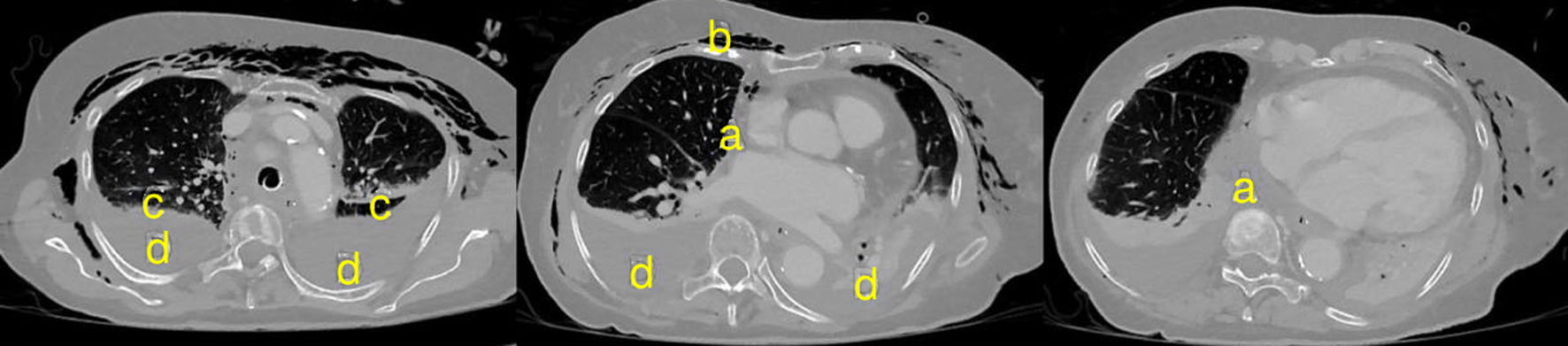 Click for large image | Figure 1. Thoracic computed tomography in an 86-year-old patient approximately 48 h after esophageal perforation caused by transesophageal echocardiography. Paraesophageal and mediastinal fluid collections (a), cervicothoracic emphysema (b), subtotal atelectasis of both lower lobes (c), and bilateral pleural effusions (d) are visible. |
Using ICU admission as the reference point because the exact time of injury was often uncertain, the first endoscopy was performed after a median of 2.0 (2.0–12.0) h; 32/41 patients (78.0%) underwent endoscopy within 12 h and 36/41 (87.8%) within 24 h. In the perforation group, 19/21 patients (90.5%) underwent endoscopy within 12 h and 20/21 (95.2%) within 24 h, compared with 13/20 (65.0%) and 16/20 (80.0%), respectively, in the group without perforation.
Supportive care was initiated within 2 h after ICU admission in all patients and comprised patient-specific combinations of nil per os, anti-infective therapy, proton pump inhibition, enteral feeding, or decompression using a triple-lumen tube, parenteral nutrition, and pleural or mediastinal drainage. Anti-infective therapy was administered to 40/42 patients (95.2%); 35/40 treated patients (87.5%) received a combination of at least two agents. Piperacillin/tazobactam was used in 25/40 (62.5%) and was combined with caspofungin in 22/25 of these cases (88.0%). Meropenem was used in 22/40 (55.0%). Bacteria or fungi were identified in 13/42 patients (31.0%) from blood, pleural fluid, sputum, bronchial secretions, urine, or perioperative specimens. The median duration of anti-infective therapy was 7 (3–17) days. A triple-lumen feeding/decompression tube was placed in 17/42 patients (40.5%).
Because treatment modalities could be combined or used sequentially, four mutually exclusive treatment patterns were identified: supportive care without defect-directed EVT or surgery in 16/42 patients (38.1%), EVT without surgery in 11/42 (26.2%), surgery without EVT in 7/42 (16.7%), and combined EVT and surgery in 8/42 (19.0%). Overall, EVT was performed in 19/42 patients (45.2%) and surgery in 15/42 (35.7%). EVT was more frequent in patients with perforation than in those without perforation (14/22 (63.6%) vs. 5/20 (25.0%); P = 0.016), as was surgery (12/22 (54.5%) vs. 3/20 (15.0%); P = 0.011). All eight patients receiving combined EVT and surgery had confirmed perforation (36.4% vs. 0.0%; P = 0.004).
Intracavitary EVT was the preferred primary endoscopic defect treatment and was performed in 19/42 patients (45.2%). Two of these 19 patients additionally received an esophageal stent in individually selected cases; thus, stent placement did not increase the total number of patients receiving defect-directed endoscopic therapy. EVT was initiated a median of 2.0 (1.5–3.0) h after ICU admission; 18/19 patients (94.7%) started EVT within 12 h, and all 19 within 16 h. The median duration of EVT was 5.0 (3.5–11.0) days.
Among the 18 patients with perforation who received EVT or surgery, the first defect-directed intervention was initiated after a median of 2.5 (1.3–4.8) h; 16/18 (88.9%) were treated within 12 h, one between 12 and 24 h, and one after 24 h. Among the eight patients without perforation who received a defect-directed intervention, the corresponding time was 2.0 (2.0–3.0) h; 7/8 (87.5%) were treated within 12 h and one between 12 and 24 h. EVT was started within 12 h in 13/14 patients with perforation and in all 5/5 patients without perforation; the remaining patient with perforation started EVT between 12 and 24 h.
Among the seven patients treated with surgery but not EVT, surgery was initiated after a median of 4.0 (2.5–17.0) h; 5/7 underwent surgery within 12 h and 6/7 within 24 h. In the eight patients receiving combined therapy, EVT preceded surgery and was initiated after 3.5 (2.0–6.0) h, whereas subsequent surgery was performed after 45.0 (32.8–69.0) h. Across all 26 patients receiving a defect-directed intervention, the first intervention occurred after 2.0 (2.0–4.0) h; 23/26 (88.5%) were treated within 12 h and 25/26 (96.2%) within 24 h.
Treatment patterns varied descriptively by lesion location. EVT and surgery were used, respectively, in 1/8 and 1/8 patients with cervical injury, 9/12 and 3/12 with thoracic injury, 3/5 and 2/5 with thoracoabdominal injury, 6/12 and 8/12 with abdominal injury, and 0/5 and 1/5 with injury involving the entire esophagus. These distributions are descriptive because treatment selection also depended on defect characteristics, contamination, tissue viability, and clinical status.
The 15 operated patients underwent a total of 28 operations (median 1; range, 1–7). Operative components included cervical diversion, exploratory thoracotomy, primary repair or oversewing, mediastinal, pleural, or cervical drainage and lavage, surgical vacuum-assisted closure procedures, and esophageal resection, including Ivor Lewis and discontinuity procedures. Several patients underwent more than one type of operation as part of sequential source-control and reconstructive strategies.
Mortality
In-hospital all-cause mortality was 2.4% (n = 1). Because only one death occurred, analyses of mortality according to perforation, empyema, sepsis, mediastinitis, peritonitis, or surgical treatment are descriptive and do not permit meaningful inference regarding mortality predictors.
Systematic post-discharge follow-up was not available. Consequently, longer-term survival, readmissions, reinterventions, dysphagia, stricture formation, nutritional status, and reconstructive outcomes could not be compared between treatment strategies.
| Discussion | ▴Top |
Clinical outcomes and perforation-associated morbidity
In this retrospective cohort of critically ill patients with non-surgical esophageal injury, confirmed perforation identified a substantially more severe clinical phenotype. Patients with perforation had more mediastinitis, pleural empyema, sepsis, septic shock, acute kidney injury, invasive ventilation, and a markedly longer ICU stay. The association between perforation and infectious complications supports its use as a clinically relevant marker of disease severity rather than as an isolated anatomical finding. In-hospital mortality was 2.4%, whereas recent population-based and systematic evidence has reported considerably higher short-term mortality in broader cohorts of esophageal perforation or trauma [8–13].
The low mortality observed in our cohort should not be interpreted as evidence of a causal benefit of any specific treatment strategy. The French nationwide cohort of 4,765 patients reported 90-day mortality of 28% and demonstrated an association between higher institutional case volume and lower mortality and failure-to-rescue [8]. Differences in etiology, outcome definitions, referral pathways, case selection, and treatment availability preclude direct comparison. Early referral, rapid access to CT and endoscopy, specialized surgical and endoscopic expertise, and survivorship bias may all have contributed to the favorable in-hospital survival in our cohort.
Diagnostics
Our findings support a low threshold for CT and endoscopy when clinically relevant esophageal injury is suspected. CT is important for localizing perforation, defining mediastinal and pleural contamination, and guiding drainage or surgical planning [3, 7, 14]. Endoscopy provides direct assessment of mucosal injury, defect dimensions, endoscopic accessibility, and tissue viability; it may also enable immediate EVT or selected stent placement. Procedural insufflation, extension of an existing defect, airway compromise, and hemodynamic instability must nevertheless be considered, particularly when full-thickness perforation is suspected [3, 14].
In our cohort, 97.6% of patients underwent endoscopy, with 78.0% examined within 12 h and 87.8% within 24 h after ICU admission. The complementary use of CT and endoscopy may reduce occult perforations because either modality alone can miss selected lesions. However, ICU admission was used as the timing anchor because onset was often uncertain; these data therefore demonstrate rapid in-center availability rather than the interval from injury to diagnosis. The retrospective design does not permit attribution of outcomes to early endoscopy.
Respiratory and critical care implications
Respiratory complications were common and multifactorial. Chemical and mechanical injury may directly affect the upper airway, whereas perforation can cause mediastinitis, pleural effusion, empyema, and secondary pulmonary dysfunction. In our cohort, invasive ventilation was more frequent in patients with perforation, although ventilation duration was similar between groups. These findings support early anticipation of airway difficulty and pulmonary complications, especially when perforation is suspected [15].
Persistent airway swelling occurred in 14.3% of patients, and tracheotomy was performed in 16.7%, broadly comparable with prior observations after caustic ingestion [15]. Pleural effusion and empyema may further complicate weaning [16]. In patients with cervical injury or planned cervical diversion, the tracheotomy strategy should be coordinated with the surgical team. Noninvasive ventilation may be considered selectively, but positive airway pressure could theoretically worsen a cervical lesion or contribute to perforation, and isolated cases have been reported [17].
Anti-infective and supportive therapy
The indication for anti-infective therapy depends on etiology, clinical presentation, and complications. A restrictive approach may be reasonable in isolated mucosal or caustic injury without pulmonary involvement, whereas broad anti-infective coverage should be considered when perforation is proven or strongly suspected. Mediastinitis and pleural empyema generally require antimicrobial therapy together with adequate source control [2, 7, 10, 15, 18, 19]. Anti-infective therapy was recorded in 40/42 patients in our cohort and was started as part of early supportive care. The frequent use of combination therapy and antifungal agents reflects center-specific practice and cannot establish superiority over narrower regimens; repeated microbiological reassessment and de-escalation remain important.
Supportive management in this cohort comprised individualized combinations of nil per os, proton pump inhibition, anti-infective treatment, enteral feeding, and decompression using a triple-lumen tube, parenteral nutrition, and pleural or mediastinal drainage [2, 4–6, 20, 21]. These measures were initiated within 2 h after ICU admission in all patients and continued alongside EVT or surgery when indicated. Accordingly, the category supportive care without defect-directed intervention denotes the absence of EVT or surgery rather than an absence of active treatment or source-control procedures.
Endoscopic and surgical management
Contemporary management is characterized by individualized and frequently multimodal strategies rather than a uniform hierarchy of conservative, endoscopic, and surgical treatment [22–26]. Intracavitary EVT was the preferred primary endoscopic defect treatment in our cohort and was used in 19 patients; two of these patients also received an esophageal stent, and eight required additional surgery. Recent multicenter data reported successful defect closure with EVT in 24/27 patients with non-postoperative esophageal perforation [27]. In a multicenter cohort of Boerhaave syndrome, primary EVT was successful in 80% of patients [28], while a 2025 meta-analysis reported a pooled clinical success rate of 88.9% for transmural perforations [29]. A multinational cohort of 216 patients with Boerhaave syndrome further demonstrated contemporary use of endoscopic, surgical, and combined strategies in 32%, 34%, and 31% of patients, respectively [30]. These non-randomized data support EVT as an important option but do not establish its superiority for all defect types or clinical presentations.
Surgery remains necessary in selected patients with large or freely communicating defects, uncontrolled contamination, severe mediastinitis or pleural empyema, tissue necrosis, clinical instability, sepsis, or failure of endoscopic treatment. Contemporary surgical data continue to show substantial morbidity despite evolving procedural choices [31, 32]. In our cohort, surgery was more frequent in patients with perforation, and operative procedures ranged from repair and drainage to diversion and resection. The eight combined EVT-surgery cases all had perforation, illustrating that endoscopic defect control and operative source control may be complementary rather than competing strategies. Because treatment assignment was strongly influenced by disease severity, comparisons of outcomes between strategies would be confounded by indication.
Pragmatic treatment algorithm
Based on the present cohort, clinical routine, and current literature, the pragmatic algorithm in Figure 2 emphasizes immediate stabilization, prompt CT and/or endoscopy, early supportive care, and repeated interdisciplinary reassessment.
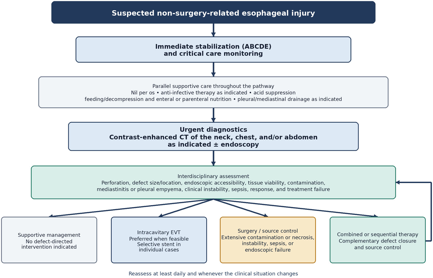 Click for large image | Figure 2. Pragmatic, non-validated treatment pathway for patients with suspected non-surgery-related esophageal injury. Supportive measures continue throughout treatment. The pathway should be reassessed at least daily and whenever the clinical situation changes. ABCDE: airway, breathing, circulation, disability, and exposure; CT: computed tomography; EVT: endoscopic vacuum therapy. |
The choice of supportive-only, endoscopic, surgical, or combined treatment should consider perforation, defect size and location, endoscopic accessibility, tissue viability, contamination, mediastinitis or pleural empyema, instability, sepsis, and response to initial therapy. No fixed algorithm was used in this cohort. When expertise or resources are insufficient, early communication with and transfer to a specialized high-volume center should be considered [8, 12, 30, 33].
Strengths and limitations
This study provides detailed information on organ dysfunction, airway and respiratory management, anti-infective therapy, diagnostic timing, and overlapping interdisciplinary treatment in a rare clinical condition. However, the retrospective single-center design, small sample size, heterogeneous etiologies, and exploratory analyses limit generalizability and preclude causal inference. Multiple statistical comparisons increase the risk of type I error, and no adjustment for multiplicity was performed. Treatment groups were not randomized and were strongly affected by disease severity and response to previous therapy, preventing comparative effectiveness conclusions. The exact time of injury was often unknown; timing from ICU admission reflects in-center processes but may underestimate the total delay from injury, especially after interhospital transfer. Referral and survivorship bias are likely, and the low number of deaths precludes meaningful analysis of mortality predictors. Finally, systematic post-discharge follow-up was unavailable, so long-term survival, reinterventions, swallowing function, strictures, nutritional recovery, and reconstruction could not be assessed.
Conclusions
Non-surgical esophageal injury in critically ill patients is rare and heterogeneous, ranging from mucosal injury to free perforation with severe mediastinal, pleural, and septic complications. Confirmed perforation was associated with greater morbidity and prolonged ICU treatment.
| Supplementary Material | ▴Top |
Suppl 1. ICD-10 codes used for patient identification.
Acknowledgments
Not applicable.
Financial Disclosure
No study-specific funding was obtained.
Conflict of Interest
DTK has a grant for an investigator-initiated study from Distalmotion. SHC has participated on a data safety monitoring or advisory board for Medtronic. HFF has participated on data safety monitoring or advisory boards for Distalmotion, Medtronic, and Stryker and holds stock or stock options in Fortimedix. CJB has participated on a data safety monitoring or advisory board for Medtronic. FD received a conference fee from the German Society of Anesthesiology and Intensive Care Medicine and study equipment from bioMérieux SA, Marcy-l’Étoile, France. The remaining authors declare no competing interests.
Informed Consent
The Ethics Committee waived the requirement for informed consent because the study was retrospective and used fully anonymized data. The radiographic figure contains no identifiable patient information.
Author Contributions
PKO: conceptualization, data curation, formal analysis, investigation, methodology, project administration, validation, visualization, and writing – original draft. DH and WAW: conceptualization and investigation. SHC, DTK, and HFF: writing – original draft. CNH: writing – review and editing. CJB and AUS: supervision and writing – review and editing. FD: supervision, conceptualization, and writing – review and editing. All authors reviewed and approved the final manuscript and agree to be accountable for all aspects of the work.
Data Availability
The datasets used and analyzed during the current study are available from the corresponding author on reasonable request.
AI Use Declaration
OpenAI ChatGPT was used during revision to assist with English-language refinement, organization of revised sections, and verification of descriptive summaries derived from the anonymized dataset. It was used in the Abstract, Introduction, Materials and Methods, Results, Discussion, Conclusions, and point-by-point response. All statistical outputs, references, interpretations, and final wording were independently checked and approved by the authors, who take full responsibility for the content.
Abbreviations
AKI: acute kidney injury; BMI: body mass index; CRP: C-reactive protein; CT: computed tomography; EVT: endoscopic vacuum therapy; HFNO: high-flow nasal oxygen; ICD: International Classification of Diseases; ICU: intensive care unit; IQR: interquartile range; NIV: noninvasive ventilation
| References | ▴Top |
- Bryant AS, Cerfolio RJ. Esophageal trauma. Thorac Surg Clin. 2007;17(1):63-72.
doi pubmed - Chirica M, Kelly MD, Siboni S, Aiolfi A, Riva CG, Asti E, Ferrari D, et al. Esophageal emergencies: WSES guidelines. World J Emerg Surg. 2019;14:26.
doi pubmed - Papaconstantinou D, Kapetanakis EI, Mylonakis A, Davakis S, Kotidis E, Tagkalos E, Rouvelas I, et al. Current aspects in the management of esophageal trauma: a systematic review and proportional meta-analysis. Dis Esophagus. 2024;37(6):doae007.
doi pubmed - Bonavina L, Chirica M, Skrobic O, Kluger Y, Andreollo NA, Contini S, Simic A, et al. Foregut caustic injuries: results of the world society of emergency surgery consensus conference. World J Emerg Surg. 2015;10:44.
doi pubmed - Kluger Y, Ishay OB, Sartelli M, Katz A, Ansaloni L, Gomez CA, Biffl W, et al. Caustic ingestion management: world society of emergency surgery preliminary survey of expert opinion. World J Emerg Surg. 2015;10:48.
doi pubmed - Schraufnagel DP, Mubashir M, Raymond DP. Non-iatrogenic esophageal trauma: a narrative review. Mediastinum. 2022;6:23.
doi pubmed - Schroder W, Leers JM, Bludau M, Herbold T, Holscher AH. [Perforations near the cardia in benign diseases]. Chirurg. 2014;85(12):1064-1072.
doi pubmed - Veziant J, Boudis F, Lenne X, Bruandet A, Eveno C, Nuytens F, Piessen G. Outcomes associated with esophageal perforation management: results from a French nationwide population-based cohort study. Ann Surg. 2023;278(5):709-716.
doi pubmed - Sdralis EIK, Petousis S, Rashid F, Lorenzi B, Charalabopoulos A. Epidemiology, diagnosis, and management of esophageal perforations: systematic review. Dis Esophagus. 2017;30(8):1-6.
doi pubmed - Navarro-Reynoso FP, Vargas-Abrego B, Pérez-Romo A, Cicero-Sabido R. Mediastinitis, a model of care. Experience in the General Hospital of Mexico over 34 years (1982-2016). Rev Med Hosp Gen Mex. 2017;80(4):228-232.
doi - Khan JA, Lehtomaki AI, Toikkanen VJ, Ukkonen MT, Nevalainen RM, Laurikka JO. Long-term prognosis and causes of death after pleural infections. Scand J Surg. 2018;107(2):145-151.
doi pubmed - Markar SR, Mackenzie H, Wiggins T, Askari A, Faiz O, Zaninotto G, Hanna GB. Management and outcomes of esophageal perforation: a national study of 2,564 patients in England. Am J Gastroenterol. 2015;110(11):1559-1566.
doi pubmed - Bayram AS, Erol MM, Melek H, Colak MA, Kermenli T, Gebitekin C. The success of surgery in the first 24 hours in patients with esophageal perforation. Eurasian J Med. 2015;47(1):41-47.
doi pubmed - Makhani M, Midani D, Goldberg A, Friedenberg FK. Pathogenesis and outcomes of traumatic injuries of the esophagus. Dis Esophagus. 2014;27(7):630-636.
doi pubmed - Struck MF, Beilicke A, Hoffmeister A, Gockel I, Gries A, Wrigge H, Bernhard M. Acute emergency care and airway management of caustic ingestion in adults: single center observational study. Scand J Trauma Resusc Emerg Med. 2016;24:45.
doi pubmed - Bediwy AS, Al-Biltagi M, Saeed NK, Bediwy HA, Elbeltagi R. Pleural effusion in critically ill patients and intensive care setting. World J Clin Cases. 2023;11(5):989-999.
doi pubmed - Van de Louw A, Brocas E, Boiteau R, Perrin-Gachadoat D, Tenaillon A. Esophageal perforation associated with noninvasive ventilation: a case report. Chest. 2002;122(5):1857-1858.
doi pubmed - Contini S, Scarpignato C. Caustic injury of the upper gastrointestinal tract: a comprehensive review. World J Gastroenterol. 2013;19(25):3918-3930.
doi pubmed - Avner BS, Ginosyan A, Le J, Mak J, Qiryaqoz Z, Huffman C. Analysis of antibiotic use and clinical outcomes in adults with known and suspected pleural empyema. BMC Infect Dis. 2022;22(1):783.
doi pubmed - Obarski P, Wlodarczyk J. Diagnosis and management of gastrointestinal chemical burns and post-burn oesophageal stenosis. Kardiochir Torakochirurgia Pol. 2021;18(4):252-259.
doi pubmed - Palmowski L, von Busch A, Unterberg M, Bergmann L, Schmitz S, Schluter A, Peters J, et al. Timely cessation of proton pump inhibitors in critically ill patients impacts morbidity and mortality: a propensity score-matched cohort study. Crit Care Med. 2024;52(2):190-199.
doi pubmed - Messager M, Warlaumont M, Renaud F, Marin H, Branche J, Piessen G, Mariette C. Recent improvements in the management of esophageal anastomotic leak after surgery for cancer. Eur J Surg Oncol. 2017;43(2):258-269.
doi pubmed - Scognamiglio P, Reeh M, Karstens K, Bellon E, Kantowski M, Schon G, Zapf A, et al. Endoscopic vacuum therapy versus stenting for postoperative esophago-enteric anastomotic leakage: systematic review and meta-analysis. Endoscopy. 2020;52(8):632-642.
doi pubmed - Plum PS, Herbold T, Berlth F, Christ H, Alakus H, Bludau M, Chang DH, et al. Outcome of self-expanding metal stents in the treatment of anastomotic leaks after Ivor Lewis esophagectomy. World J Surg. 2019;43(3):862-869.
doi pubmed - Kamarajah SK, Bundred J, Spence G, Kennedy A, Dasari BVM, Griffiths EA. Critical appraisal of the impact of oesophageal stents in the management of oesophageal anastomotic leaks and benign oesophageal perforations: an updated systematic review. World J Surg. 2020;44(4):1173-1189.
doi pubmed - Berlth F, Bludau M, Plum PS, Herbold T, Christ H, Alakus H, Kleinert R, et al. Self-Expanding Metal Stents Versus Endoscopic Vacuum Therapy in Anastomotic Leak Treatment After Oncologic Gastroesophageal Surgery. J Gastrointest Surg. 2019;23(1):67-75.
doi pubmed - Luttikhold J, Pattynama LMD, Seewald S, Groth S, Morell BK, Gutschow CA, Ida S, et al. Endoscopic vacuum therapy for esophageal perforation: a multicenter retrospective cohort study. Endoscopy. 2023;55(9):859-864.
doi pubmed - Wannhoff A, Kouladouros K, Koschny R, Walter B, Zoll Z, Buringer K, Blank S, et al. Endoscopic vacuum therapy for the treatment of Boerhaave syndrome: a multicenter analysis. Gastrointest Endosc. 2025;101(2):365-374.
doi pubmed - Vohra I, Gopakumar H, Sharma NR, Puli SR. Efficacy of endoscopic vacuum therapy in esophageal luminal defects: a systematic review and meta-analysis. Clin Endosc. 2025;58(1):53-62.
doi pubmed - Hauge T, Abu Hejleh A, Aiolfi A, Berlth F, Bonavina L, Brake L, Conradi LC, et al. Treatment strategies for Boerhaave syndrome: multinational retrospective cohort study. Br J Surg. 2025;112(12):znaf260.
doi pubmed - Axtell AL, Gaissert HA, Morse CR, Premkumar A, Schumacher L, Muniappan A, Ott H, et al. Management and outcomes of esophageal perforation. Dis Esophagus. 2022;35(1):1-8.
doi pubmed - Wong LY, Leipzig M, Liou DZ, Backhus LM, Lui NS, Shrager JB, Berry MF. Surgical management of esophageal perforation: examining trends in a multi-institutional cohort. J Gastrointest Surg. 2023;27(9):1757-1765.
doi pubmed - Thim T, Krarup NH, Grove EL, Rohde CV, Lofgren B. Initial assessment and treatment with the Airway, Breathing, Circulation, Disability, Exposure (ABCDE) approach. Int J Gen Med. 2012;5:117-121.
doi pubmed
This article is distributed under the terms of the Creative Commons Attribution 4.0 International License (CC BY 4.0), which permits unrestricted use, distribution, and reproduction in any medium, including commercial use, provided the original work is properly cited.
Gastroenterology Research is published by Elmer Press Inc.