


Localized Gastric Amyloid Light Chain Amyloidosis With Atypical Depressed Morphology: Diagnostic Implications Based on Magnifying Endoscopy
DOI:
https://doi.org/10.14740/gr2176Keywords:
Narrow-band imaging, Early gastric cancer, Lymphoma, StomachAbstract
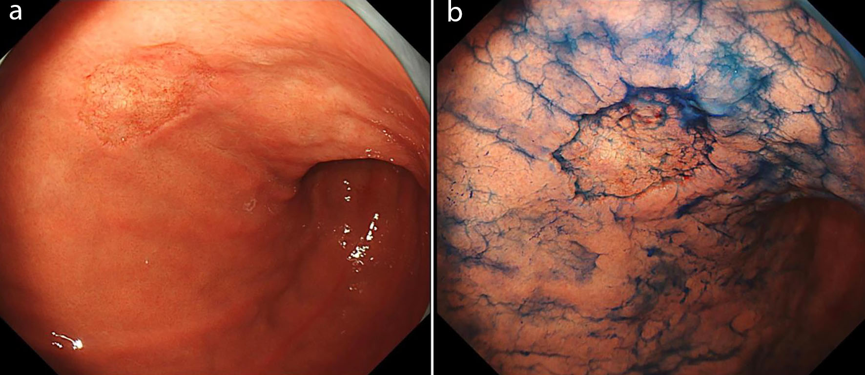
Gastrointestinal involvement of amyloidosis is uncommon and cases limited to the stomach with a depressed lesion are limited. Here, we report the case of a 78-year-old woman who had a solitary 25-mm depressed lesion with irregular and indistinct boundaries in the lower gastric body with esophagogastroduodenoscopy (EGD). Chromoendoscopy (CE) revealed a coarse mucosal surface with a “moth-eaten” appearance, suggesting early gastric cancer. Magnifying narrow-band imaging (NBI) revealed partially preserved glandular structures and tortuous vessels without definite irregular microvascular patterns (IMVPs). Endoscopic ultrasonography (EUS) revealed a homogeneous hypoechoic thickening confined to the superficial layers. Histopathological examination revealed amyloid deposits in the submucosa. Congo red staining was positive and immunohistochemical analysis confirmed amyloid light-chain lambda-type (AL-λ) deposition. Comprehensive systemic evaluation showed no evidence of systemic involvement, establishing a diagnosis of localized gastric immunoglobulin light chain amyloidosis. Further accumulation of similar cases is required to clarify the characteristic endoscopic findings and their clinical significance.
Published
Issue
Section
License
Copyright (c) 2026 The authors

This work is licensed under a Creative Commons Attribution 4.0 International License.