| Gastroenterology Research, ISSN 1918-2805 print, 1918-2813 online, Open Access |
| Article copyright, the authors; Journal compilation copyright, Gastroenterol Res and Elmer Press Inc |
| Journal website https://gr.elmerpub.com |
Original Article
Volume 19, Number 5, October 2026, pages 273-287

Systems Biology and Machine Learning Reveal Oxidative Stress Hubs and Therapeutic Targets in Diarrhea-Predominant Irritable Bowel Syndrome
Shu Fei Zhanga, c, Wei Wei Xieb, c, Yu Xin Lia, Yu Qian Zhangb, Yi Ran Jinb, d, Ying Feng Dua, d
aDepartment of Pharmaceutical Analysis, School of Pharmacy, Hebei Medical University, Shijiazhuang, Hebei 050017, China.
bDepartment of Clinical Pharmacy, The Second Hospital of Hebei Medical University, Shijiazhuang, Hebei 050000, China
cThese authors contributed equally to this work.
dCorresponding Authors: Yi Ran Jin, Department of Clinical Pharmacy, The Second Hospital of Hebei Medical University, Shijiazhuang, Hebei 050000, China; Ying Feng Du, Department of Pharmaceutical Analysis, School of Pharmacy, Hebei Medical University, Shijiazhuang, Hebei 050017, China
Manuscript submitted May 28, 2026, accepted August 21, 2026, published online October 31, 2026
Short title: Oxidative Stress Hubs in IBS-D
doi: https://doi.org/10.14740/gr2172
| Abstract | ▴Top |
Background: Diarrhea-predominant irritable bowel syndrome (IBS-D) is a common functional gastrointestinal disorder, and accumulating evidence suggests that oxidative stress is involved in its pathophysiology. This study explored oxidative stress–related mechanisms in IBS-D and screened potential diagnostic biomarkers and therapeutic targets through integrative bioinformatics analysis.
Methods: Transcriptomic datasets from the National Center for Biotechnology Information (NCBI)-Gene Expression Omnibus (GEO) database were analyzed using integrative bioinformatics approaches to identify oxidative stress–related core genes in IBS-D. Eight potential biomarkers were identified and evaluated for diagnostic performance via receiver operating characteristic (ROC) analysis. Among these, the key gene ACO2 was further investigated using Kyoto Encyclopedia of Genes and Genomes (KEGG)-based gene set enrichment analysis (GSEA) and immune correlation analyses to explore the association between ACO2 expression and disease-related biological processes. In addition, the eight target proteins were used for reverse screening of potentially therapeutic traditional Chinese medicines and their active compounds.
Results: In total, 206 oxidative stress–related genes were identified, with predominant enrichment observed in mitochondrial energy metabolism and cellular stress–response pathways, including oxidative phosphorylation and hypoxia-inducible factor 1 (HIF-1) signaling. The eight potential diagnostic biomarkers demonstrated robust diagnostic performance (area under the ROC curve (AUC) = 0.896) in distinguishing IBS-D patients from healthy controls. Notably, ACO2 emerged as a key hub gene associated with distinct metabolic and immune-related pathway enrichment profiles. In parallel, a competing endogenous RNA (ceRNA) regulatory network centered on ACO2 was established, providing additional insight into its potential post-transcriptional regulatory mechanisms. Furthermore, reverse screening based on the eight target proteins identified luteolin, phillygenin, quercetin, and wogonin as candidate active compounds from Forsythia suspensa. Molecular docking further indicated favorable binding affinity between these compounds and the biomarker proteins.
Conclusions: This integrative bioinformatics study identifies oxidative stress–related molecular signatures and potential biomarkers in IBS-D. Reverse screening and molecular docking further suggest that luteolin, phillygenin, quercetin, and wogonin from Forsythia suspensa may represent potential candidate compounds targeting these biomarkers. These findings provide a theoretical basis for further experimental validation.
Keywords: Oxidative stress; Machine-learning algorithm; Hub gene; Immune infiltration
| Introduction | ▴Top |
Irritable bowel syndrome (IBS) is a common functional heterogeneous gut-brain interaction disorder characterized by complex, multifactorial mechanisms [1]. Clinically, it primarily manifests as abdominal pain, bloating, and altered bowel habits, along with a range of physiological and psychological disturbances. Based on predominant bowel habits, IBS is generally classified into four subtypes: constipation-predominant, diarrhea-predominant, mixed, and unclassified IBS [2]. Affecting approximately 11% of the global population [3], IBS has no definitive cure and significantly impairs quality of life, potentially leading to substantial disability [4]. At present, the exact etiology of diarrhea-predominant irritable bowel syndrome (IBS-D) remains unclear. Multiple factors—including intestinal barrier dysfunction, dysregulation of neural signaling, genetic factors, and stress—have been increasingly implicated in its pathogenesis [5–8]. Meanwhile, oxidative stress and inflammatory responses are critically involved in the pathogenesis of IBS-D [9–11]. Inflammation may exacerbate IBS-D symptoms by activating visceral sensory pathways and disrupting gastrointestinal physiological reflexes [12, 13]. It can also amplify pain perception by promoting the release of pro-inflammatory mediators, such as cytokines, which activate intracellular signaling pathways and enhance neural sensitivity [14, 15].
Visceral hypersensitivity, resulting from inflammation and tissue injury, is thought to underlie the abdominal discomfort and pain in patients with IBS [16]. Relative to healthy controls, IBS patients exhibit altered cytokine profiles, characterized by increased plasma concentrations of interleukin (IL)-6 and IL-8, indicating low-grade inflammatory and immune responses. These cytokines may further affect the hypothalamus, enhancing stress responses [17, 18]. The hypothalamic–pituitary–adrenal (HPA) axis is closely involved in neuroendocrine regulation under stress conditions. Pro-inflammatory cytokines, such as IL-6 and IL-8, can stimulate the release of corticotropin-releasing hormone (CRH), thereby modulating intestinal immune function [9, 19–21].
Oxidative stress is commonly defined as an imbalance between reactive oxygen species (ROS) production and antioxidant clearance, and this process is closely involved in inflammatory responses [22, 23]. Excessive ROS generation can induce cellular damage and activate inflammation-related transcription factors, thereby triggering inflammatory responses [24–26]. These processes may compromise intestinal epithelial integrity, leading to increased intestinal permeability and enhanced mucosal immune activation, ultimately exacerbating disease progression in patients [27–29]. While oxidative stress and inflammatory responses are well established features of inflammatory bowel disease [30–32], their molecular signatures and regulatory networks in IBS-D remain insufficiently characterized. Therefore, the present study aimed to prioritize oxidative stress–related candidate biomarkers in IBS-D through integrative bioinformatics and machine-learning analyses, and to predict traditional Chinese medicine–derived bioactive compounds for future experimental validation.
| Materials and Methods | ▴Top |
Gene set collection and pre-processing
Datasets GSE36701 and GSE166869 were retrieved from the National Center for Biotechnology Information (NCBI)-Gene Expression Omnibus (GEO) database. As a training dataset for discovery, the GSE36701 dataset is constructed from the GPL570 platform. In this microarray study, the cohort consisted of 27 patients with IBS-D and 19 healthy controls, from whom rectal mucosal biopsy samples were obtained. The preprocessed series matrix file (GSE36701_series_matrix) was downloaded from GEO and used for analysis. Probe annotation was conducted using the corresponding platform annotation file (GPL570). Probe identifiers were matched to official gene symbols, and expression values of multiple probes corresponding to the same gene were averaged. For individuals with multiple biopsies, one representative sample per subject was retained to avoid pseudo-replication. For validation, we utilized the gene expression dataset GSE166869, which comprises duodenal-jejunal biopsy samples from 26 healthy controls and 23 patients.
Screening of IBS-D–associated genes
Differentially expressed genes (DEGs) between IBS-D and control samples in the GSE36701 dataset were identified using the R package limma, with |log2 fold change| ≥ 0.585 and adjusted P < 0.05 as the screening criteria [33]. In parallel, weighted gene coexpression network analysis (WGCNA) was performed to construct gene coexpression networks and identify modules associated with IBS-D [34].
Prior to network construction, probe-level expression values were collapsed to gene-level values, and genes with low expression variability across samples were filtered out (standard deviation ≤ 0.1). Sample clustering was first performed to assess sample quality and identify potential outliers. Samples exceeding the hierarchical clustering cut height of 20,000 were removed from subsequent analyses. An unsigned weighted gene coexpression network was then constructed. The optimal soft-thresholding power was selected according to the scale-free topology criterion, and a value of 8 was chosen to approximate scale-free network characteristics. Subsequently, the adjacency matrix was transformed into a TOM-based dissimilarity matrix, which served as the input for hierarchical clustering. Gene modules were then defined by the dynamic tree cutting method, with parameters configured as a minimum cluster size of 60 and a deepSplit of 2. Modules with high similarity were subsequently merged using a cut height of 0.25 to generate the final module structure. Finally, the correlations between genes and modules (MM) as well as modules and traits (GS) are calculated to assess the significance and importance of each module.
IBS-D oxidative stress–related genes
We queried the GeneCards database for oxidative stress–associated genes, applying a relevance score threshold of > 7 to enrich for high-confidence candidates potentially linked to IBS-D. Subsequently, the three gene lists—DEGs, WGCNA module genes, and OS_genes—were intersected to obtain the overlapping candidates for further analysis. The chromosomal locations of the identified IBS-D–related oxidative stress genes were then annotated using the “circlize” package (version 0.4.16), and a Manhattan plot was generated using the “CMplot” package (version 4.5.1) to visualize their genomic distribution.
To further explore the biological roles of these overlapping genes, functional enrichment analyses were performed using the R package clusterProfiler. Enriched terms and pathways with both P value and adjusted P value < 0.05 were considered statistically significant.
Construction of protein–protein interaction (PPI) network analysis
We used the STRING database to build a protein–protein interaction (PPI) network and examine the interrelationships among the oxidative stress–related genes. After excluding free unconnected proteins [35], a PPI network was constructed and visualized in Cytoscape (version 3.10.0) [36–38]. Then, the cytoHubba (version 0.1), a module identification plugin in Cytoscape (version 3.10.0), was used to perform topological analysis based on three algorithms: maximal neighborhood component (MNC), Degree, and edge percolated component (EPC). For each algorithm, the top 20 ranked genes were selected, and the overlapping genes among the three algorithms were defined as hub genes, resulting in a total of 10 hub genes.
To further characterize the biological relevance of the identified hub genes, differential expression analysis, gene–gene correlation analysis, and gene–immune cell association analysis were performed. Differences in the expression levels of the 10 hub genes between groups were assessed and displayed using box plot visualization. Pairwise correlations among core genes were assessed using Spearman correlation analysis, and the relationships were visualized using scatter plots and correlation heatmaps. Only gene pairs with a Spearman correlation coefficient greater than 0.4 in absolute value and a P value < 0.05 were retained for visualization. In addition, immune cell infiltration in IBS-D samples was evaluated using ssGSEA, and Spearman correlation analysis was performed to explore the relationships between the 10 hub genes and immune cell infiltration scores, with the results presented as lollipop plots.
Identification of IBS-D potential biomarkers
To identify oxidative stress–related potential diagnostic biomarkers associated with IBS-D, a least absolute shrinkage and selection operator (LASSO) regression analysis was conducted using the R package glmnet (version 4.1.8). LASSO is a penalized regression method that introduces an L1 regularization term to shrink regression coefficients, thereby enabling feature selection and reducing the risk of overfitting [39]. A binomial logistic regression (LASSO) model with α = 1 was fitted using the gene expression data as predictors and disease status as the binary response. The optimal tuning parameter λ was selected by 10-fold cross-validation, with λ_min defined as the value yielding the minimum cross-validated deviance. Genes with nonzero regression coefficients at λ_min were retained for further evaluation as potential biomarkers.
Subsequently, receiver operating characteristic (ROC) curve analysis was carried out using the R package pROC (version 1.18.5) to assess the diagnostic value of biomarkers selected by the LASSO model. Model discrimination was assessed using the area under the ROC curve (AUC), with the 95% confidence interval (CI) estimated by 2,000 bootstrap resampling iterations.
Intergroup analysis
The signature gene with the highest score identified from the PPI network constructed in Section 2.4 was selected for further in-depth investigation. To explore pathway-level differences associated with this gene, all samples were stratified into high- and low-expression groups based on the median expression value of the target gene. For gene set enrichment analysis (GSEA), genes were ranked according to their log fold change (logFC) values between the two expression groups. GSEA was conducted with the “clusterProfiler” package, and pathways with P < 0.05 were considered significantly enriched. In addition, gene set variation analysis (GSVA) based on Kyoto Encyclopedia of Genes and Genomes (KEGG) gene sets was used to assess pathway-level differences between the high- and low-expression groups, with statistical significance determined by a t-test at P < 0.05.
To further explore the relationship between the signature gene and immune functional activity, immune function scores were computed for each sample using predefined immune-related gene sets. Differences in immune functional activity between high- and low-expression groups were subsequently evaluated, and statistically significant immune functions associated with differential gene expression were identified.
ceRNA network construction
To systematically investigate potential regulatory relationships among the signature gene, we performed the following analyses. First, putative human miRNA–mRNA interactions (hsa) were identified through integrated data mining of four established databases: miRDB [40] (version 6.0), miRanda [41] (version 3.3), TargetScan [42] (version 8.0), and spongeScan (version 1.1). Only microRNAs (miRNAs) predicted to target the core genes by at least three of these databases were retained for subsequent analysis. Using the spongeScan [43] computational tool, we then predicted long noncoding RNAs (lncRNAs) capable of interacting with these selected miRNAs. Finally, these interaction datasets were integrated to construct a comprehensive miRNA–lncRNA–mRNA regulatory network, which was visualized using Cytoscape (v3.10.0).
Prediction of candidate herbal active compounds
To further predict oxidative stress–related active herbal compounds with potential therapeutic effects on IBS-D, the eight candidate therapeutic target genes were first standardized using the UniProt database. The standardized gene symbols were then imported into the Traditional Chinese Medicine Systems Pharmacology Database and Analysis Platform (TCMSP), HERB, and BATMAN-TCM databases to retrieve compounds potentially interacting with these targets. Active compounds obtained from the TCMSP database were further screened according to the criteria of oral bioavailability (OB) ≥ 30% and drug-likeness (DL) ≥ 0.18. The screened compounds were then integrated and subjected to enrichment analysis, with pvalueFilter = 0.05 and adj PvalFilter = 0.05 set as the filtering thresholds. Finally, the predicted active herbal compounds were selected for molecular docking analysis with the potential diagnostic target proteins. The two-dimensional (2D) structures of the candidate compounds were obtained from the PubChem database, while the three-dimensional (3D) structures of the target proteins were retrieved from the Protein Data Bank (Supplementary Material 1, gr.elmerpub.com). Molecular docking was performed using AutoDock Vina [44], and the docking results were subsequently visualized and analyzed using Discovery Studio 4.5.
Institutional Review Board approval and ethical compliance
Institutional Review Board approval and ethical compliance approval were not required for this study because it did not involve new human participants, animal experiments, or patient-identifiable information.
| Results | ▴Top |
Integrated screening of IBS-D-related genes
First, analysis of the GSE36701 expression dataset yielded 3,037 DEGs, and a volcano plot was generated to illustrate their overall distribution (Fig. 1a). In parallel, WGCNA was conducted using the gene expression matrix to obtain modules associated with IBS-D. The soft-thresholding power was set to 8, where the scale-free topology fit index reached 0.9 (Fig. 1c). Based on hierarchical clustering, 11 gene modules were obtained. Module–trait correlation analysis showed that the turquoise module had the strongest association with IBS-D (Cor = 0.97, P < 0.05), and contained 7,913 genes (Fig. 1b, d). The turquoise module was therefore selected as the candidate module for further analysis.
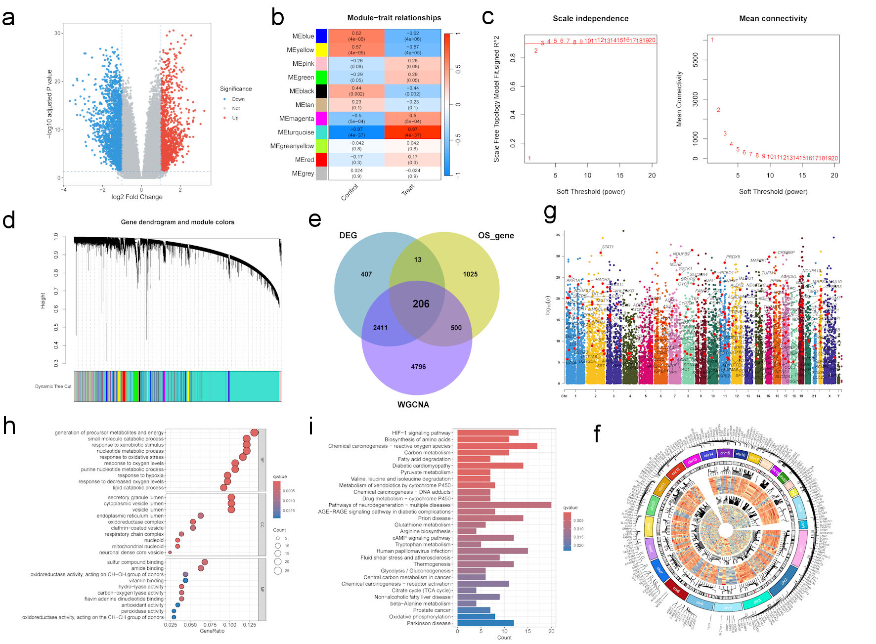 Click for large image | Figure 1. Integrated identification and functional characterization of oxidative stress–related differentially expressed genes in IBS-D. (a) Volcano plot of DEGs in the GSE36701 dataset. (b–d) WGCNA analysis of identifying candidate genes. (b) The clustered modules of WGCNA. (c) The soft threshold power and the mean connectivity of WGCNA. (d) The cluster dendrogram of WGCNA. (e) Venn diagram showing the intersection among DEGs, oxidative stress–related genes, and genes from clinically relevant WGCNA modules. (f) Circos plot showing the chromosomal locations of the oxidative stress–related DEGs. (g) Manhattan plot displaying the chromosomal significance distribution of DEGs. (h, i) Enrichment analysis of oxidative stress–related DEGs. (h) GO enrichment analysis. (i) KEGG enrichment analysis. DEGs: differentially expressed genes; GO: Gene Ontology; IBS-D: diarrhea-predominant irritable bowel syndrome; KEGG: Kyoto Encyclopedia of Genes and Genomes; WGCNA: weighted gene coexpression network analysis. |
Screening of oxidative stress–related genes in IBS-D
A total of 1744 oxidative stress–related genes were obtained from the GeneCards database (version 5.22) using a relevance score greater than 7 as the screening condition. A total of 206 oxidative stress–related genes were obtained by subjecting DEGs, oxidative stress–related genes, and genes within the module to Venn analysis (Fig. 1e).
The chromosomal distribution of these genes was subsequently analyzed. A circular heat map (Fig. 1f) illustrates the genomic locations of oxidative stress–related genes across different chromosomes, along with their expression patterns and clustering relationships. A Manhattan-like plot (Fig. 1g) was further constructed to visualize gene-level differential significance across chromosomes. Among these genes, STAT1, signal transducer and activator of transcription 1 (STAT1) gene on chromosome 2 showed notable significance. Given its central role in the Janus kinase–signal transducer and activator of transcription (JAK–STAT) signaling pathway, altered STAT1 expression may be involved in IBS-D pathogenesis by modulating immune and inflammatory responses.
Functional enrichment analysis
The Gene Ontology (GO) enrichment analysis results (Fig. 1h) revealed that, within the biological process category, the genes were predominantly enriched in pathways associated with energy metabolism and catabolic processes, including generation of precursor metabolites and energy and lipid catabolic process. In terms of cellular components, the enriched genes were mainly localized to vesicle-associated structures and organelles, including secretory granule lumen, cytoplasmic vesicle lumen and endoplasmic reticulum lumen. Regarding molecular functions (MF), the genes were significantly enriched in oxidoreductase activity, antioxidant activity, and peroxidase activity.
KEGG pathway analysis indicated that these genes were primarily associated with oxidative stress and mitochondrial energy metabolism, including hypoxia-inducible factor 1 (HIF-1) signaling, ROS-related processes, and oxidative phosphorylation (Fig. 1i). Notably, these potential genes may contribute to disease progression by regulating key biological processes such as energy metabolism, antioxidant defense, and hypoxia adaptation.
Hub gene expression and immune correlation analysis
To explore the functional interactions among the 206 intersecting genes, a PPI network was established through the STRING database and visualized in Cytoscape. Subsequent topology analysis of this network yielded 10 hub genes (Fig. 2). To further characterize the expression patterns and interrelationships of these hub genes, their expression levels were compared between groups and visualized using box plots. As shown in (Supplementary Material 2A–J, gr.elmerpub.com), all 10 genes were significantly differentially expressed between the two groups. In addition, multiple hub gene pairs exhibited significant positive correlations (Supplementary Material 2K–R, gr.elmerpub.com), suggesting a coordinated expression tendency among these genes.
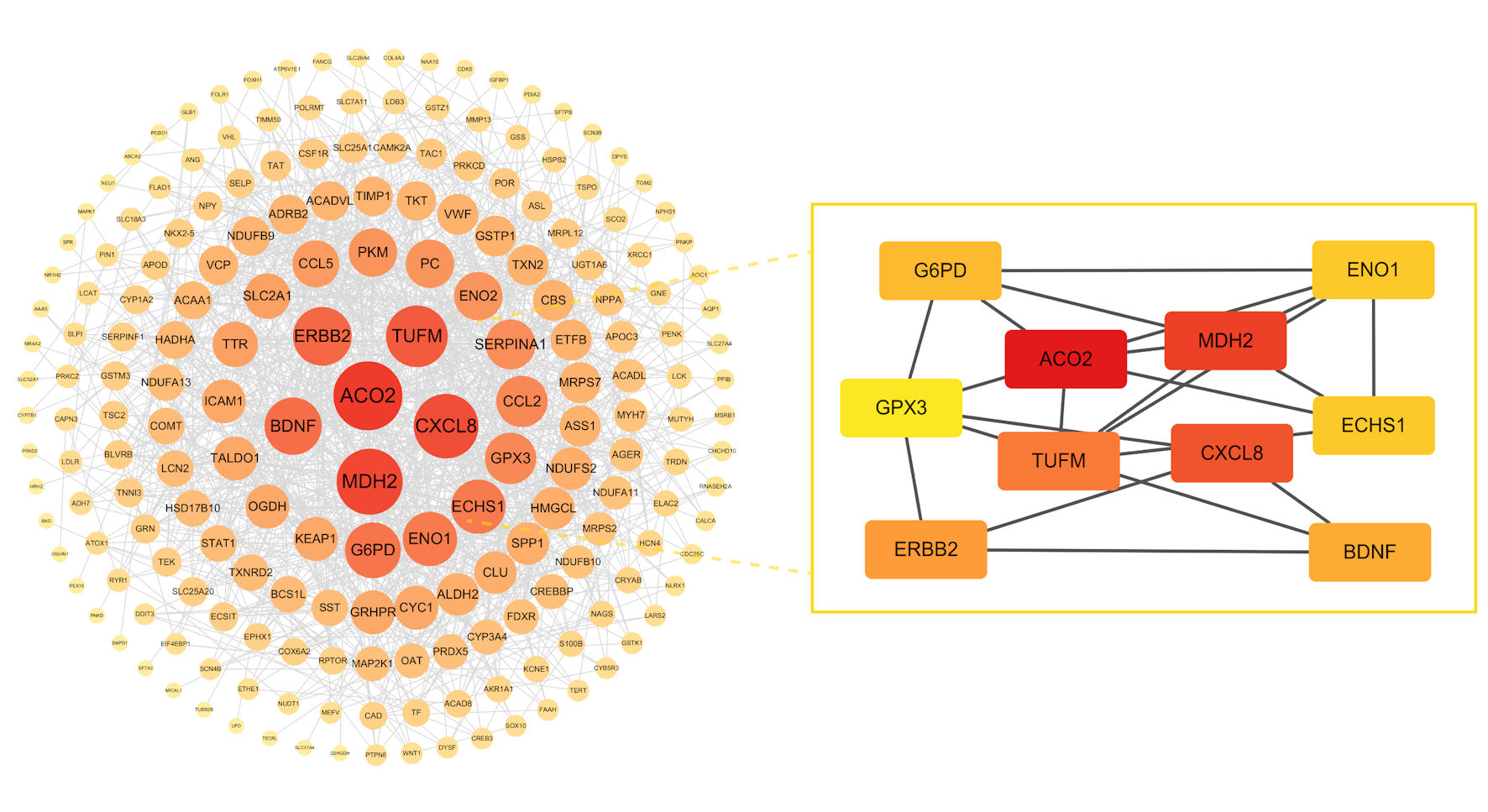 Click for large image | Figure 2. Construction of protein–protein interaction (PPI) network and identification of 10 hub genes. |
We next examined the relationships between the 10 hub genes and immune cell infiltration scores in IBS-D samples. Notably, three genes—ACO2, BDNF, and ERBB2—showed significant correlations with specific immune cell types (Fig. 3a–c), suggesting that these genes may be involved in the regulation of the immune microenvironment.
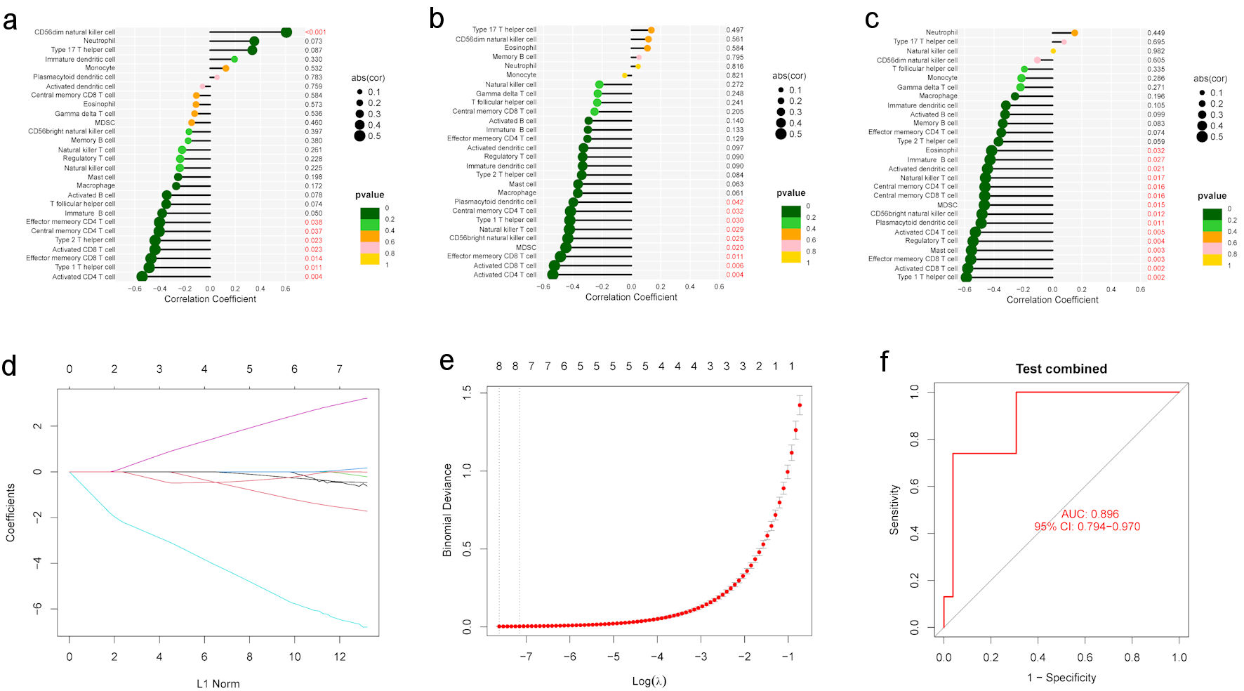 Click for large image | Figure 3. Immune cell correlation analysis and construction of a diagnostic model. (a–c) Correlation between hub genes and infiltrating immune cells. (a) Correlation between ACO2 and infiltrating immune cells. (b) Correlation between BDNF and infiltrating immune cells. (c) Correlation between ERBB2 and infiltrating immune cells. (d, e) The LASSO regression process for feature selection. (d) LASSO path plot. (e) LASSO cross-validation error plot. (f) Diagnostic performance of the eight core targets in distinguishing normal and IBS-D samples, as evaluated by receiver operating characteristic (ROC) analysis in the GSE166869 dataset. LASSO: least absolute shrinkage and selection operator; IBS-D: diarrhea-predominant irritable bowel syndrome. |
Identification of eight candidate diagnostic biomarkers
Subsequently, to further evaluate the diagnostic potential of the 10 hub genes, we applied the LASSO regression (Fig. 3d, e) algorithm with 10-fold cross-validation [45]. This analysis ultimately identified eight potential diagnostic biomarkers—ECHS1, ERBB2, GPX3, CXCL8, MDH2, BDNF, ACO2, and TUFM—with significant diagnostic value for further investigation (Table 1). Based on these genes, ROC curve analysis was performed in the validation cohort GSE166869. As shown in Figure 3f, the model achieved an AUC of 0.896 (95% CI, 0.791–0.973), indicating strong discriminative ability for distinguishing IBS-D samples from normal controls.
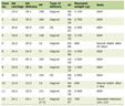 Click to view | Table 1. List of Identified Potential Biomarkers and Their Regulation |
Pathway enrichment stratified by ACO2
We also performed GSEA based on KEGG pathways to identify ACO2-related signaling pathways in IBS-D (Fig. 4a, b). As shown in Figure 4a, metabolic pathways closely related to mitochondrial function and oxidative stress, including the tricarboxylic acid (TCA) cycle and arginine and proline metabolism, were predominantly enriched in the ACO2 high-expression group, whereas immune-related pathways, such as cytokine–cytokine receptor interaction and chemokine signaling, were mainly enriched in the ACO2 low-expression group.
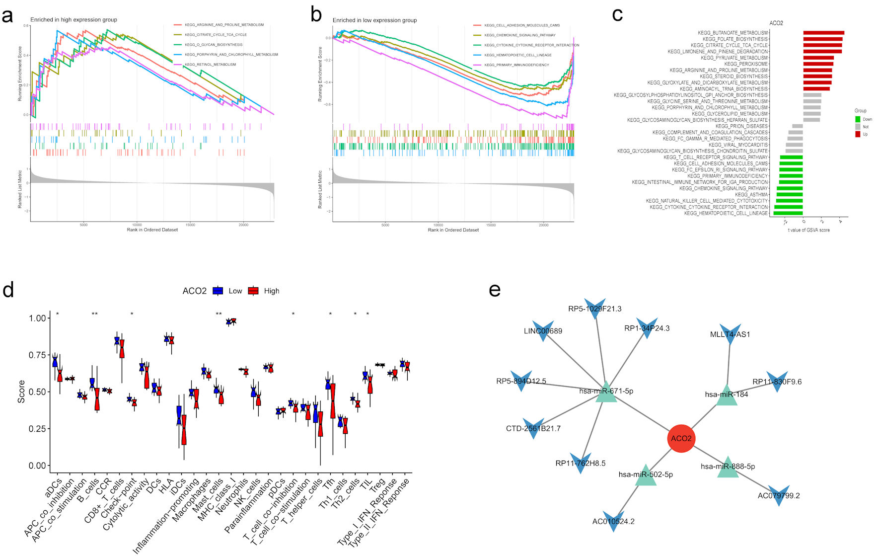 Click for large image | Figure 4. Functional enrichment analysis and immune landscape associated with ACO2 expression in IBS-D. (a, b) Gene set enrichment analysis. (a) GSEA enrichment results of ACO2 high expression group. (b) GSEA enrichment results of ACO2 low expression group. (c) Gene set variation analysis (GSVA) of differential pathway enrichment between groups. (d) Comparison of ssGSEA Immune score among ACO2 high- and low-expression groups. (e) The ceRNA network illustrating the interactions among ACO2, related miRNAs, and lncRNAs. IBS-D: diarrhea-predominant irritable bowel syndrome; GSEA: gene set enrichment analysis; GSVA: gene set variation analysis; ssGSEA: single-sample gene set enrichment analysis; ceRNA: competing endogenous RNA; lincRNAs: long noncoding RNAs. |
In parallel, differential pathway activation analysis using GSVA further revealed distinct functional activation patterns between the two groups (Fig. 4c). Pathways potentially activated in the ACO2 high-expression group were mainly related to energy metabolism, amino acid metabolism, and peroxisomal function, which may reflect enhanced mitochondrial metabolic activity. In contrast, the ACO2 low-expression group showed significant enrichment in pathways linked to immune and inflammatory responses, including T-cell receptor signaling, NK cell–mediated cytotoxicity, and intestinal immune regulation.
Immune function differences associated with target gene expression
The immune cell–associated activity of the high- and low-expression groups was further analyzed to evaluate differences in the immune microenvironment. As shown in Figure 4d, significant differences in immune functional activity were observed between the high- and low-ACO2 expression groups. Notably, the ACO2 low-expression group exhibited significantly higher levels of multiple immune-related functions compared with the high-expression group, including activated dendritic cells (aDCs), B cells, immune checkpoint activity, mast cells, T-cell co-inhibition, and Th2 cells. These results suggest that reduced ACO2 expression is associated with enhanced immune-related activity in IBS-D. Consistently, GSVA analysis showed preferential activation of immune-related pathways in the ACO2 low-expression group, further supporting a potential link between ACO2 downregulation and immune dysregulation.
ceRNA network construction
To reveal the regulatory mechanism of target gene expression, this study constructed a ceRNA network of target genes to analyze the mechanism of miRNA and lncRNA regulation of the ACO2 gene (Fig. 4e). The miRNAs and lncRNAs of target genes compete through common miRNA binding sites, which in turn regulate the post-transcriptional regulation level of target genes. In the constructed network diagram, the target gene is regulated through four miRNAs, and each miRNA in turn interacts with multiple lncRNAs separately, resulting in the final regulatory network diagram. Specifically, the lncRNAs affect the expression of the target gene by competitively binding to the miRNAs, thereby influencing their binding to the target mRNA. In-depth analysis of these interactions can provide new perspectives for understanding the regulatory mechanisms in specific biological processes of target genes. The complete list of interactions was provided here (Supplementary Material 3, gr.elmerpub.com).
Candidate active herbal compound screening and molecular docking
At the initial stage of candidate drug screening, the names of the target proteins were first standardized using the UniProt database. Compounds potentially interacting with these target proteins were then retrospectively screened from multiple databases, resulting in 151 compound–target interaction pairs (Supplementary Material 4, gr.elmerpub.com). Subsequent enrichment analysis showed that Forsythia suspensa was enriched with six active compounds, representing the highest number of enriched compounds among the identified traditional Chinese medicines (Supplementary Material 5, gr.elmerpub.com). Among these compounds, quercetin, luteolin, wogonin, and phillygenin were identified as the major candidate compounds because they were associated with multiple target proteins (Table 2).
Click to view | Table 2. Active Compounds of Forsythia suspensa Identified by Enrichment Analysis |
Therefore, these four compounds were selected for molecular docking analysis. The docking results showed that the selected compounds exhibited favorable binding affinities with the target proteins, with binding energies ranging from −10.0 to −6.7 kcal/mol (Fig. 5a). All ligand–protein complexes formed more than two hydrogen bonds within the binding pockets. Notably, the aconitase 2/aconitate hydratase (ACO2)–phillygenin and erb-b2 receptor tyrosine kinase 2 (ERBB2)–luteolin complexes each formed five hydrogen bonds, indicating stable molecular interactions between the candidate compounds and their corresponding target proteins (Fig. 5b–f; Supplementary Material 6, gr.elmerpub.com).
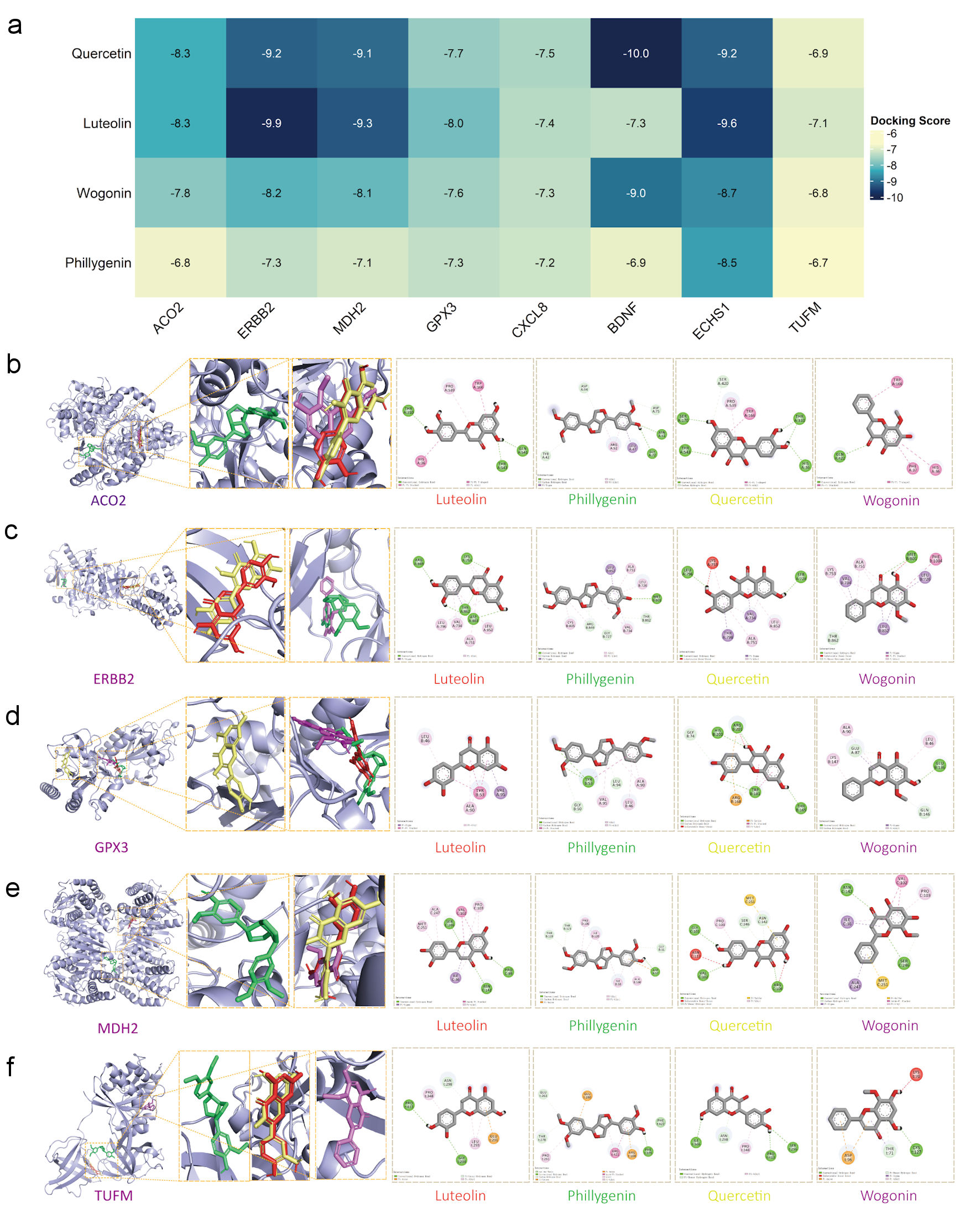 Click for large image | Figure 5. Prediction of interactions between potential active compounds and core proteins by molecular docking analysis. (a) Heatmap of molecular docking binding energies. Lower binding energies represented by yellow modules and higher binding energies represented by blue modules. (b–f) Structural diagrams of active compound–target protein complexes. The magnified three-dimensional (3D) diagrams show the binding sites of the four compounds within the protein pockets, while the two-dimensional (2D) diagrams illustrate the intermolecular interaction bonds between the compounds and target proteins. ACO2: aconitase 2/aconitate hydratase; BDNF: brain-derived neurotrophic factor; ERBB2: erb-b2 receptor tyrosine kinase 2; GPX3: glutathione peroxidase 3; MDH2: malate dehydrogenase 2; TUFM: Tu translation elongation factor, mitochondrial. |
| Discussion | ▴Top |
IBS-D is a common functional gastrointestinal disorder with a multifactorial and incompletely understood pathogenesis. Although multiple mechanisms—including oxidative stress, mitochondrial dysfunction, immune dysregulation, and barrier impairment—have been investigated in IBS-D, their molecular interconnections remain unclear [9, 46–50]. Here, we applied an integrative bioinformatics and network pharmacology approach to characterize oxidative stress–related molecular signatures in IBS-D, prioritize candidate biomarkers and therapeutically relevant targets, and predict active compounds from traditional Chinese medicines for further validation.
Specifically, by integrating differential expression analysis, WGCNA, protein interaction analysis, and LASSO regression, we identified eight oxidative stress–related hub genes with good diagnostic performance in an independent dataset (AUC = 0.896). Considering the multi-target therapeutic characteristics of traditional Chinese medicine, we further performed reverse pharmacology screening and molecular docking to prioritize candidate herbal compounds for future validation.
Previous studies have implicated brain-derived neurotrophic factor (BDNF) in gut–brain axis dysfunction and visceral hypersensitivity in IBS-D, partly through the BDNF/tropomyosin receptor kinase B (TrkB)/phospholipase C gamma 1 (PLCγ1) signaling pathway [51–53]. Clinical evidence has further shown that serum BDNF and proBDNF levels were elevated in children with IBS and positively associated with symptom severity, particularly abdominal pain and visceral hypersensitivity [54]. Beyond its neuromodulatory role, BDNF may also participate in mucosal neuro–immune interactions by modulating immune cell activation and inflammatory signaling [55]. Given that BDNF expression and signaling are sensitive to oxidative stress–related microenvironmental changes [56, 57], BDNF may serve as a molecular link connecting oxidative stress, immune activation, and neural sensitization in IBS-D, thereby supporting its potential diagnostic relevance. CXCL8, also known as IL-8, is a member of the CXC chemokine family that promotes immune cell recruitment and inflammatory responses through the CXCR1/CXCR2 axis and downstream MAPK, PI3K-Akt, and NF-κB signaling pathways [58]. This is consistent with the recognized role of low-grade inflammation and immune activation in IBS-D pathophysiology. Previous studies have reported elevated IL-8 levels in patients with IBS-D, which were negatively correlated with occludin expression [59]. These findings suggest that CXCL8-mediated inflammatory cell recruitment and persistent low-grade inflammation may further compromise intestinal epithelial barrier integrity, thereby increasing intestinal permeability. Therefore, CXCL8 may contribute to IBS-D through an inflammation–barrier impairment axis and may represent a candidate biomarker and potential therapeutic target.
In addition, enoyl-CoA hydratase short-chain 1 (ECHS1), malate dehydrogenase 2 (MDH2), ACO2, and Tu translation elongation factor, mitochondrial (TUFM) are closely associated with mitochondrial function and energy metabolism [60–63]. Although direct experimental evidence linking these genes to IBS-D remains limited, their biological functions are highly consistent with key pathophysiological features of IBS-D, including impaired mucosal energy metabolism, mitochondrial dysfunction, and disrupted ROS homeostasis. Previous proteomic studies have reported abnormal expression of multiple energy metabolism-related proteins in the colonic mucosa of patients with IBS-D, accompanied by reduced ATP levels, suggesting that mucosal energy metabolic dysregulation may contribute to IBS-D pathogenesis [50]. In parallel, glutathione peroxidase 3 (GPX3) and ERBB2 may be closely associated with redox-related dysregulation in IBS-D, although they likely act through different biological mechanisms. As an extracellular glutathione peroxidase involved in antioxidant defense, GPX3 may reflect impaired antioxidant defenses or reduced ROS-scavenging capacity in IBS-D [64]. From a gastrointestinal perspective, insufficient glutathione-dependent antioxidant protection may increase mucosal susceptibility to oxidative injury, promote lipid peroxidation, and impair epithelial barrier integrity [31, 65]. Indeed, oxidative stress has been reported to disrupt epithelial tight junctions through redox-sensitive modifications of junctional proteins, while extracellular glutathione peroxidase has been shown to be involved in intestinal inflammatory responses [66, 67]. Therefore, altered GPX3 expression may not only reflect redox imbalance but also contribute to the interaction between oxidative stress, barrier dysfunction, and mucosal inflammation in IBS-D. ERBB2 encodes a receptor tyrosine kinase, and its downstream signaling has been implicated in cellular responses to oxidative stress and redox-sensitive regulatory networks [68]. Previous studies have reported that ErbB2 overexpression can increase glutathione peroxidase activity and reduce basal ROS levels [69], suggesting that ERBB2 may be involved in redox-sensitive signaling regulation. Clinically, excessive ROS production has been suggested to contribute to diarrhea-related intestinal dysfunction [70, 71]. Collectively, these findings support the hypothesis that the candidate genes identified in this study may participate in IBS-D pathophysiology through the convergence of mitochondrial dysfunction, oxidative stress, and immune-inflammatory activation.
Given its central position in the PPI network, ACO2 was further investigated as a potential hub gene linking mitochondrial metabolic dysfunction with immune dysregulation in IBS-D. ACO2 encodes mitochondrial aconitase, a key enzyme in the TCA cycle that is essential for ATP production and metabolic homeostasis [72, 73]. In the present study, reduced ACO2 expression was observed in IBS-D samples and was associated with increased immune infiltration, suggesting that impaired mitochondrial metabolic activity may be accompanied by a more inflammatory mucosal microenvironment. This finding is biologically plausible because mitochondrial aconitase contains an iron–sulfur cluster that is highly sensitive to ROS-mediated oxidation, and oxidative stress may further compromise aconitase activity [74]. Therefore, reduced ACO2 expression, together with the potential oxidative inactivation of mitochondrial aconitase, may reflect a compromised TCA cycle and impaired mucosal energy metabolism in IBS-D. Notably, TCA cycle intermediates are not merely metabolic substrates but can also act as signaling molecules that regulate immune cell activation and inflammatory responses [75, 76]. In addition, previous studies have shown that mitochondrial aconitase activity can modulate immune function by altering oxaloacetate levels and activating mitochondrial stress signaling pathways [77]. Therefore, the observed association between low ACO2 expression and enhanced immune infiltration may reflect a potential metabolic–immune interaction in IBS-D.
Reverse pharmacology screening provides a useful target-driven strategy for linking disease-associated molecular targets with potential bioactive compounds by integrating target information with pharmacological databases. In the present study, among the predicted traditional Chinese medicine–derived compounds, quercetin, luteolin, wogonin, and phillygenin were prioritized because they were associated with multiple target proteins, suggesting their potential multi-target regulatory capacity. In addition, molecular docking analysis revealed favorable binding energies between these compounds and their corresponding target proteins, indicating that they may have the capacity to interact with key molecules involved in oxidative stress–related dysregulation in IBS-D. Therefore, these compounds should be regarded as promising candidate molecules for future experimental validation.
Several limitations of the present study should nevertheless be considered. This study was conducted solely based on bioinformatics analyses and lacked experimental validation in vivo and in vitro, which may limit mechanistic interpretation. Moreover, the relatively small sample size may affect the robustness of the conclusions. Importantly, the training cohort (GSE36701) was derived from rectal mucosal biopsies, whereas the external validation cohort (GSE166869) originated from duodenal–jejunal biopsies. This cross-site validation introduces biological heterogeneity due to regional differences in intestinal function and gene expression; therefore, the validation may reflect both IBS-D–related signals and inter-segmental variation. Nevertheless, the observed validation performance may also imply that the identified candidate diagnostic biomarkers represent relatively stable molecular alterations across different intestinal regions. In addition, the screening of active herbal compounds was based on computational prediction and pharmacological databases, and the potential interactions between candidate compounds and target proteins remain to be experimentally confirmed. Therefore, further studies integrating functional assays, IBS-D animal models, and pharmacological validation are needed to confirm the biological relevance, diagnostic value, and clinical utility of the identified candidate biomarkers, as well as the pharmacological potential of the predicted bioactive compounds.
Conclusions
In conclusion, this study applied an integrative bioinformatics and machine-learning framework to systematically characterize oxidative stress–related molecular features in IBS-D. We identified a panel of candidate oxidative stress–associated biomarkers with potential diagnostic value, which were potentially associated with mitochondrial dysfunction, immune infiltration, metabolic dysregulation and gut–brain axis dysregulation in IBS-D. Furthermore, reverse screening based on the predicted candidate targets identified several candidate bioactive compounds derived from traditional Chinese medicine, including quercetin, luteolin, phillygenin, and wogonin, providing preliminary computational evidence for their potential therapeutic relevance in IBS-D. Overall, these findings provide computational evidence for oxidative stress–related molecular alterations in IBS-D and offer candidate biomarkers, targets, and compounds for future experimental validation.
| Supplementary Material | ▴Top |
Suppl 1. Details of core target proteins used for molecular docking analysis.
Suppl 2. Box plots showing the differential expression of hub genes and scatter plots illustrating the correlations among hub genes.
Suppl 3. Predicted human (hsa) miRNA–lncRNA–mRNA interactions associated with core genes.
Suppl 4. Target–compound associations between core proteins and potential active compounds.
Suppl 5. Traditional Chinese medicine enrichment results based on core targets.
Suppl 6. Structural diagrams of active compound–target protein complexes.
Acknowledgments
None to declare.
Financial Disclosure
This work was supported by the Hebei Traditional Chinese Medicine Administration Scientific Research Projects (Nos. 2025307 and 2025308) and the Natural Science Foundation of Hebei Province, China (No. H2024206249).
Conflict of Interest
The authors declare that there are no competing financial interests or personal relationships that could have influenced the work reported in this paper.
Informed Consent
Not applicable.
Author Contributions
Shu Fei Zhang and Wei Wei Xie performed data collection, bioinformatics analyses, and manuscript writing. Yu Xin Li and Yu Qian Zhang contributed to software and tool development for data analysis. Yi Ran Jin and Ying Feng Du designed and supervised the study. All authors read and approved the final manuscript.
Data Availability
Any inquiries regarding supporting data availability of this study should be directed to the corresponding author.
Abbreviations
ACO2: aconitase 2/aconitate hydratase; aDCs: activated dendritic cells; AUC: area under the receiver operating characteristic curve; BDNF: brain-derived neurotrophic factor; ceRNA: competing endogenous RNA; CI: confidence interval; CRH: corticotropin-releasing hormone; DEGs: differentially expressed genes; DL: drug-likeness; ECHS1: enoyl-CoA hydratase short-chain 1; EPC: edge percolated component; MNC: maximal neighborhood component; GEO: Gene Expression Omnibus; GO: Gene Ontology; GPX3: glutathione peroxidase 3; GS: gene significance; GSEA: gene set enrichment analysis; GSVA: gene set variation analysis; HIF-1: hypoxia-inducible factor 1; HPA: hypothalamic–pituitary–adrenal; IBS: irritable bowel syndrome; IBS-D: diarrhea-predominant irritable bowel syndrome; IL: interleukin; JAK–STAT: Janus kinase–signal transducer and activator of transcription; KEGG: Kyoto Encyclopedia of Genes and Genomes; LASSO: least absolute shrinkage and selection operator; MDH2: malate dehydrogenase 2; MM: module membership; NCBI: National Center for Biotechnology Information; NF-κB: nuclear factor kappa B; Nrf2: nuclear factor erythroid 2-related factor 2; OB: oral bioavailability; PDB: Protein Data Bank; PLCγ1: phospholipase C gamma 1; PPI: protein–protein interaction; RCSB: Research Collaboratory for Structural Bioinformatics; ROC: receiver operating characteristic; ROS: reactive oxygen species; ssGSEA: single-sample gene set enrichment analysis; STAT1: signal transducer and activator of transcription 1; TCA: tricarboxylic acid; TCMSP: Traditional Chinese Medicine Systems Pharmacology Database and Analysis Platform; TrkB: tropomyosin receptor kinase B; TUFM: Tu translation elongation factor, mitochondrial; WGCNA: weighted gene coexpression network analysis
| References | ▴Top |
- Biesiekierski JR, Manning LP, Murray HB, Vlaeyen JWS, Ljotsson B, Van Oudenhove L. Review article: exclude or expose? The paradox of conceptually opposite treatments for irritable bowel syndrome. Aliment Pharmacol Ther. 2022;56(4):592-605.
doi pubmed - Enck P, Aziz Q, Barbara G, Farmer AD, Fukudo S, Mayer EA, Niesler B, et al. Irritable bowel syndrome. Nat Rev Dis Primers. 2016;2:16014.
doi pubmed - Lovell RM, Ford AC. Global prevalence of and risk factors for irritable bowel syndrome: a meta-analysis. Clin Gastroenterol Hepatol. 2012;10(7):712-721.e714.
doi pubmed - Holtmann GJ, Ford AC, Talley NJ. Pathophysiology of irritable bowel syndrome. Lancet Gastroenterol Hepatol. 2016;1(2):133-146.
doi pubmed - Balmus IM, Lefter R, Ciobica A, Cojocaru S, Guenne S, Timofte D, Stanciu C, et al. Preliminary biochemical description of brain oxidative stress status in irritable bowel syndrome contention-stress rat model. Medicina (Kaunas). 2019;55(12):776.
doi pubmed - Nam SY, Kim BC, Ryu KH, Park BJ. Prevalence and risk factors of irritable bowel syndrome in healthy screenee undergoing colonoscopy and laboratory tests. J Neurogastroenterol Motil. 2010;16(1):47-51.
doi pubmed - Qin HY, Cheng CW, Tang XD, Bian ZX. Impact of psychological stress on irritable bowel syndrome. World J Gastroenterol. 2014;20(39):14126-14131.
doi pubmed - Lu Y, Chai Y, Qiu J, Zhang J, Wu M, Fu Z, Wang Y, et al. Integrated omics analysis reveals the epigenetic mechanism of visceral hypersensitivity in IBS-D. Front Pharmacol. 2023;14:1062630.
doi pubmed - Choghakhori R, Abbasnezhad A, Hasanvand A, Amani R. Inflammatory cytokines and oxidative stress biomarkers in irritable bowel syndrome: association with digestive symptoms and quality of life. Cytokine. 2017;93:34-43.
doi pubmed - Zhang Y, Huang S, Zhang S, Hao Z, Shen J. Pomegranate peel extract mitigates diarrhea-predominant irritable bowel syndromes via MAPK and NF-kappaB pathway modulation in rats. Nutrients. 2024;16(22):3854.
doi pubmed - Qiao R, Sheng C, Lu Y, Zhang Y, Ren H, Lemos B. Microplastics induce intestinal inflammation, oxidative stress, and disorders of metabolome and microbiome in zebrafish. Sci Total Environ. 2019;662:246-253.
doi pubmed - Yuan Y, Wang X, Huang S, Wang H, Shen G. Low-level inflammation, immunity, and brain-gut axis in IBS: unraveling the complex relationships. Gut Microbes. 2023;15(2):2263209.
doi pubmed - Sinagra E, Pompei G, Tomasello G, Cappello F, Morreale GC, Amvrosiadis G, Rossi F, et al. Inflammation in irritable bowel syndrome: Myth or new treatment target? World J Gastroenterol. 2016;22(7):2242-2255.
doi pubmed - Sun C, Zhang J, Chen L, Liu T, Xu G, Li C, Yuan W, et al. IL-17 contributed to the neuropathic pain following peripheral nerve injury by promoting astrocyte proliferation and secretion of proinflammatory cytokines. Mol Med Rep. 2017;15(1):89-96.
doi pubmed - Ng QX, Soh AYS, Loke W, Lim DY, Yeo WS. The role of inflammation in irritable bowel syndrome (IBS). J Inflamm Res. 2018;11:345-349.
doi pubmed - Spiller R, Aziz Q, Creed F, Emmanuel A, Houghton L, Hungin P, Jones R, et al. Guidelines on the irritable bowel syndrome: mechanisms and practical management. Gut. 2007;56(12):1770-1798.
doi pubmed - Clarke G, Quigley EM, Cryan JF, Dinan TG. Irritable bowel syndrome: towards biomarker identification. Trends Mol Med. 2009;15(10):478-489.
doi pubmed - Scully P, McKernan DP, Keohane J, Groeger D, Shanahan F, Dinan TG, Quigley EM. Plasma cytokine profiles in females with irritable bowel syndrome and extra-intestinal co-morbidity. Am J Gastroenterol. 2010;105(10):2235-2243.
doi pubmed - Shulman RJ, Jarrett ME, Cain KC, Broussard EK, Heitkemper MM. Associations among gut permeability, inflammatory markers, and symptoms in patients with irritable bowel syndrome. J Gastroenterol. 2014;49(11):1467-1476.
doi pubmed - Chen Q, Ren Y, Lu J, Bartlett M, Chen L, Zhang Y, Guo X, et al. A novel prebiotic blend product prevents irritable bowel syndrome in mice by improving gut microbiota and modulating immune response. Nutrients. 2017;9(12):1341.
doi pubmed - O'Brien R, Buckley MM, O'Malley D. Divergent effects of exendin-4 and interleukin-6 on rat colonic secretory and contractile activity are associated with changes in regional vagal afferent signaling. Neurogastroenterol Motil. 2021;33(10):e14160.
doi pubmed - Hussain T, Tan B, Yin Y, Blachier F, Tossou MC, Rahu N. Oxidative stress and inflammation: What polyphenols can do for us? Oxid Med Cell Longev. 2016;2016:7432797.
doi pubmed - Salim S. Oxidative stress and psychological disorders. Curr Neuropharmacol. 2014;12(2):140-147.
doi pubmed - Jin Q, Liu T, Qiao Y, Liu D, Yang L, Mao H, Ma F, et al. Oxidative stress and inflammation in diabetic nephropathy: role of polyphenols. Front Immunol. 2023;14:1185317.
doi pubmed - Deng Z, Li D, Wang L, Lan J, Wang J, Ma Y. Activation of GABA(B)R attenuates intestinal inflammation by reducing oxidative stress through modulating the TLR4/MyD88/NLRP3 pathway and gut microbiota abundance. Antioxidants (Basel). 2024;13(9):1141.
doi pubmed - Wang Y, Zhang J, Zhang B, Lu M, Ma J, Liu Z, Huang J, et al. Modified Gegen Qinlian decoction ameliorated ulcerative colitis by attenuating inflammation and oxidative stress and enhancing intestinal barrier function in vivo and in vitro. J Ethnopharmacol. 2023;313:116538.
doi pubmed - Sahoo DK, Heilmann RM, Paital B, Patel A, Yadav VK, Wong D, Jergens AE. Oxidative stress, hormones, and effects of natural antioxidants on intestinal inflammation in inflammatory bowel disease. Front Endocrinol (Lausanne). 2023;14:1217165.
doi pubmed - Di Vincenzo F, Del Gaudio A, Petito V, Lopetuso LR, Scaldaferri F. Gut microbiota, intestinal permeability, and systemic inflammation: a narrative review. Intern Emerg Med. 2024;19(2):275-293.
doi pubmed - Wu XX, Huang XL, Chen RR, Li T, Ye HJ, Xie W, Huang ZM, et al. Paeoniflorin prevents intestinal barrier disruption and inhibits lipopolysaccharide (LPS)-induced inflammation in Caco-2 cell monolayers. Inflammation. 2019;42(6):2215-2225.
doi pubmed - Zhang J, Cen L, Zhang X, Tang C, Chen Y, Zhang Y, Yu M, et al. MPST deficiency promotes intestinal epithelial cell apoptosis and aggravates inflammatory bowel disease via AKT. Redox Biol. 2022;56:102469.
doi pubmed - Tratensek A, Locatelli I, Grabnar I, Drobne D, Vovk T. Oxidative stress-related biomarkers as promising indicators of inflammatory bowel disease activity: a systematic review and meta-analysis. Redox Biol. 2024;77:103380.
doi pubmed - Bourgonje AR, Feelisch M, Faber KN, Pasch A, Dijkstra G, van Goor H. Oxidative stress and redox-modulating therapeutics in inflammatory bowel disease. Trends Mol Med. 2020;26(11):1034-1046.
doi pubmed - Ritchie ME, Phipson B, Wu D, Hu Y, Law CW, Shi W, Smyth GK. limma powers differential expression analyses for RNA-sequencing and microarray studies. Nucleic Acids Res. 2015;43(7):e47.
doi pubmed - Langfelder P, Horvath S. WGCNA: an R package for weighted correlation network analysis. BMC Bioinformatics. 2008;9:559.
doi pubmed - Szklarczyk D, Gable AL, Nastou KC, Lyon D, Kirsch R, Pyysalo S, Doncheva NT, et al. The STRING database in 2021: customizable protein-protein networks, and functional characterization of user-uploaded gene/measurement sets. Nucleic Acids Res. 2021;49(D1):D605-D612.
doi pubmed - Shannon P, Markiel A, Ozier O, Baliga NS, Wang JT, Ramage D, Amin N, et al. Cytoscape: a software environment for integrated models of biomolecular interaction networks. Genome Res. 2003;13(11):2498-2504.
doi pubmed - Xia J, Benner MJ, Hancock RE. NetworkAnalyst—integrative approaches for protein-protein interaction network analysis and visual exploration. Nucleic Acids Res. 2014;42(Web Server issue):W167-174.
doi pubmed - Xia J, Gill EE, Hancock RE. NetworkAnalyst for statistical, visual and network-based meta-analysis of gene expression data. Nat Protoc. 2015;10(6):823-844.
doi pubmed - Tibshirani R. Regression shrinkage and selection via the lasso. J R Stat Soc: B (Methodol). 1996;58:267-288.
doi - Chen Y, Wang X. miRDB: an online database for prediction of functional microRNA targets. Nucleic Acids Res. 2020;48(D1):D127-D131.
doi pubmed - Enright AJ, John B, Gaul U, Tuschl T, Sander C, Marks DS. MicroRNA targets in Drosophila. Genome Biol. 2003;5(1):R1.
doi pubmed - Lewis BP, Burge CB, Bartel DP. Conserved seed pairing, often flanked by adenosines, indicates that thousands of human genes are microRNA targets. Cell. 2005;120(1):15-20.
doi pubmed - Furio-Tari P, Tarazona S, Gabaldon T, Enright AJ, Conesa A. spongeScan: A web for detecting microRNA binding elements in lncRNA sequences. Nucleic Acids Res. 2016;44(W1):W176-180.
doi pubmed - Eberhardt J, Santos-Martins D, Tillack AF, Forli S. AutoDock Vina 1.2.0: new docking methods, expanded force field, and python bindings. J Chem Inf Model. 2021;61(8):3891-3898.
doi pubmed - Friedman J, Hastie T, Tibshirani R. Regularization paths for generalized linear models via coordinate descent. J Stat Softw. 2010;33(1):1-22.
pubmed - Morales-Guzman AS, Alarcon-Aguilar A, Luna-Lopez A, Santana-Vargas AD, Motola-Kuba M, Librado-Osorio R, Garcia-Alvarez JA, et al. Abnormal homeostasis in the redox state and related signaling pathways, in irritable bowel syndrome. Neurogastroenterol Motil. 2025;37(12):e70097.
doi pubmed - Nasab SJ, Feizi A, Hajihashemi P, Entezari MH, Sharma M, Adibi P, Bagherniya M. Correction: Effects of Spirulina (Arthrospira) platensis supplementation on intestinal permeability, oxidative stress markers, quality of life, and disease severity in patients with constipation-predominant irritable bowel syndrome: a randomized double-blind, placebo-controlled trial. Nutr J. 2025;24(1):83.
doi pubmed - Xia Y, Tan W, Yuan F, Lin M, Luo H. Luteolin attenuates oxidative stress and colonic hypermobility in water avoidance stress rats by activating the Nrf2 signaling pathway. Mol Nutr Food Res. 2024;68(1):e2300126.
doi pubmed - Matricon J, Meleine M, Gelot A, Piche T, Dapoigny M, Muller E, Ardid D. Review article: Associations between immune activation, intestinal permeability and the irritable bowel syndrome. Aliment Pharmacol Ther. 2012;36(11-12):1009-1031.
doi pubmed - Zhang CY, Yao X, Sun G, Yang YS. Close association between abnormal expressed enzymes of energy metabolism and diarrhea-predominant irritable bowel syndrome. Chin Med J (Engl). 2019;132(2):135-144.
doi pubmed - Fan F, Tang Y, Dai H, Cao Y, Sun P, Chen Y, Chen A, et al. Blockade of BDNF signalling attenuates chronic visceral hypersensitivity in an IBS-like rat model. Eur J Pain. 2020;24(4):839-850.
doi pubmed - Wang P, Du C, Chen FX, Li CQ, Yu YB, Han T, Akhtar S, et al. BDNF contributes to IBS-like colonic hypersensitivity via activating the enteroglia-nerve unit. Sci Rep. 2016;6:20320.
doi pubmed - Liang R, Ge W, Song X, Wang H, Cui W, Zhang X, Wei Z, et al. Paeoniflorin alleviates anxiety and visceral hypersensitivity via HPA axis and BDNF/TrkB/PLCgamma1 pathway in maternal separation-induced IBS-like rats. Curr Mol Pharmacol. 2024;17:e18761429280572.
doi pubmed - Senkal E, Durankus F, Cam S, Potas N, Beyazyuz M, Albayrak Y. Increased serum BDNF and ProBDNF levels in children with irritable bowel syndrome. J Psychosom Res. 2020;137:110216.
doi pubmed - Zhang M, Wu X, Gao H, Zhang L, Li Y, Li M, Zhao C, et al. Chinese herbal medicine for irritable bowel syndrome: a perspective of local immune actions. Am J Chin Med. 2024;52(7):2079-2106.
doi pubmed - Hsu YC, Huang YY, Tsai SY, Kuo YW, Lin JH, Ho HH, Chen JF, et al. Efficacy of probiotic supplements on brain-derived neurotrophic factor, inflammatory biomarkers, oxidative stress and cognitive function in patients with Alzheimer's dementia: a 12-week randomized, double-blind active-controlled study. Nutrients. 2023;16(1):16.
doi pubmed - Mashayekhpour MA, Hajizadeh Moghaddam A, Abouhosseini Tabari M, Ranjbar M. Neuroprotective effects of nanophytosome-encapsulated citral in a mouse model of chronic unpredictable mild stress through the suppression of oxidative stress and inflammation. Biomed Pharmacother. 2025;189:118280.
doi pubmed - Zhu Y, Yang S, Zhao N, Liu C, Zhang F, Guo Y, Liu H. CXCL8 chemokine in ulcerative colitis. Biomed Pharmacother. 2021;138:111427.
doi pubmed - Zhen Y, Chu C, Zhou S, Qi M, Shu R. Imbalance of tumor necrosis factor-alpha, interleukin-8 and interleukin-10 production evokes barrier dysfunction, severe abdominal symptoms and psychological disorders in patients with irritable bowel syndrome-associated diarrhea. Mol Med Rep. 2015;12(4):5239-5245.
doi pubmed - Fu Q, Qiu R, Li S, Qin Y, Lu Z, Liyao S, Yang Z, et al. ECHS1: pathogenic mechanisms, experimental models, and emerging therapeutic strategies. Orphanet J Rare Dis. 2025;20(1):430.
doi pubmed - Pissas G, Tziastoudi M, Divani M, Poulianiti C, Konsta MAP, Lykotsetas E, Liakopoulos V, et al. Malate dehydrogenase-2 inhibition shields renal tubular epithelial cells from anoxia-reoxygenation injury by reducing reactive oxygen species. J Biochem Mol Toxicol. 2024;38(10):e23854.
doi pubmed - Tong WH, Rouault TA. In-Gel Activity Assay of Mammalian Mitochondrial and Cytosolic Aconitases, Surrogate Markers of Compartment-Specific Oxidative Stress and Iron Status. Bio Protoc. 2024;14(23):e5126.
doi pubmed - Hershkovitz T, Kurolap A, Gonzaga-Jauregui C, Paperna T, Mory A, Wolf SE, Regeneron Genetics C, et al. A novel TUFM homozygous variant in a child with mitochondrial cardiomyopathy expands the phenotype of combined oxidative phosphorylation deficiency 4. J Hum Genet. 2019;64(6):589-595.
doi pubmed - Li L, Lu M, Peng Y, Huang J, Tang X, Chen J, Li J, et al. Oxidatively stressed extracellular microenvironment drives fibroblast activation and kidney fibrosis. Redox Biol. 2023;67:102868.
doi pubmed - Mayr L, Grabherr F, Schwarzler J, Reitmeier I, Sommer F, Gehmacher T, Niederreiter L, et al. Dietary lipids fuel GPX4-restricted enteritis resembling Crohn's disease. Nat Commun. 2020;11(1):1775.
doi pubmed - Escuder-Rodriguez JJ, Liang D, Jiang X, Sinicrope FA. Ferroptosis: biology and role in gastrointestinal disease. Gastroenterology. 2024;167(2):231-249.
doi pubmed - Barrett CW, Ning W, Chen X, Smith JJ, Washington MK, Hill KE, Coburn LA, et al. Tumor suppressor function of the plasma glutathione peroxidase gpx3 in colitis-associated carcinoma. Cancer Res. 2013;73(3):1245-1255.
doi pubmed - Gordon LI, Burke MA, Singh AT, Prachand S, Lieberman ED, Sun L, Naik TJ, et al. Blockade of the erbB2 receptor induces cardiomyocyte death through mitochondrial and reactive oxygen species-dependent pathways. J Biol Chem. 2009;284(4):2080-2087.
doi pubmed - Belmonte F, Das S, Sysa-Shah P, Sivakumaran V, Stanley B, Guo X, Paolocci N, et al. ErbB2 overexpression upregulates antioxidant enzymes, reduces basal levels of reactive oxygen species, and protects against doxorubicin cardiotoxicity. Am J Physiol Heart Circ Physiol. 2015;309(8):H1271-1280.
doi pubmed - Kruidenier L, Verspaget HW. Review article: oxidative stress as a pathogenic factor in inflammatory bowel disease—radicals or ridiculous? Aliment Pharmacol Ther. 2002;16(12):1997-2015.
doi pubmed - Bhattacharyya A, Chattopadhyay R, Mitra S, Crowe SE. Oxidative stress: an essential factor in the pathogenesis of gastrointestinal mucosal diseases. Physiol Rev. 2014;94(2):329-354.
doi pubmed - Chen Y, Cai GH, Xia B, Wang X, Zhang CC, Xie BC, Shi XC, et al. Mitochondrial aconitase controls adipogenesis through mediation of cellular ATP production. FASEB J. 2020;34(5):6688-6702.
doi pubmed - Ciccarone F, De Falco P, Ciriolo MR. Aconitase 2 sensitizes MCF-7 cells to cisplatin eliciting p53-mediated apoptosis in a ROS-dependent manner. Biochem Pharmacol. 2020;180:114202.
doi pubmed - Mansilla S, Tortora V, Pignataro F, Sastre S, Castro I, Chiribao ML, Robello C, et al. Redox sensitive human mitochondrial aconitase and its interaction with frataxin: In vitro and in silico studies confirm that it takes two to tango. Free Radic Biol Med. 2023;197:71-84.
doi pubmed - Xie A, Brunner JS, Chakraborty S, Montero AM, Bridgeman AE, Paras KI, Cui R, et al. Citrate clearance is a major function of aconitase 2 in the canonical TCA cycle. Cell. 2026;189(9):2684-2699.e2621.
doi pubmed - Martinez-Reyes I, Chandel NS. Mitochondrial TCA cycle metabolites control physiology and disease. Nat Commun. 2020;11(1):102.
doi pubmed - Kim E, Annibal A, Lee Y, Park HH, Ham S, Jeong DE, Kim Y, et al. Mitochondrial aconitase suppresses immunity by modulating oxaloacetate and the mitochondrial unfolded protein response. Nat Commun. 2023;14(1):3716.
doi pubmed
This article is distributed under the terms of the Creative Commons Attribution 4.0 International License (CC BY 4.0), which permits unrestricted use, distribution, and reproduction in any medium, including commercial use, provided the original work is properly cited.
Gastroenterology Research is published by Elmer Press Inc.