Computed Tomography-Based Assessment of Sarcopenia and Disease Progression in Pancreatic Ductal Adenocarcinoma: A Radiomics and Machine Learning Approach
DOI:
https://doi.org/10.14740/gr2132Keywords:
Sarcopenia, Pancreatic cancer, CT radiomics, Machine learning, Tumor progressionAbstract
Background: Sarcopenia is a known negative prognostic factor in oncology and is frequently observed in patients with pancreatic ductal adenocarcinoma (PDAC). Computed tomography (CT) enables longitudinal muscle assessment and may provide additional prognostic information. This study aims to assess their association with prognosis in pancreatic cancer and explore the diagnostic and prognostic value of CT radiomics.
Methods: A retrospective single-center study included 62 patients with primary PDAC who underwent at least three abdominal CT scans: baseline (t0), 3 months (t1), 6 months (t2), and, in 35 patients, 12 months (t3). CT-based sarcopenia was assessed using the psoas muscle index (PMI) based on reference cutoffs and cohort-specific sex-specific quartiles. Skeletal muscles at the L3 level were semi-automatically segmented. Radiomic features of the psoas were extracted and analyzed using k-nearest neighbor, decision tree, and random forest models. Prognostic relevance was evaluated using logistic regression and feature selection via least absolute shrinkage and selection operator (LASSO) regression. Tumor progression was assessed radiologically according to RECIST 1.1 criteria.
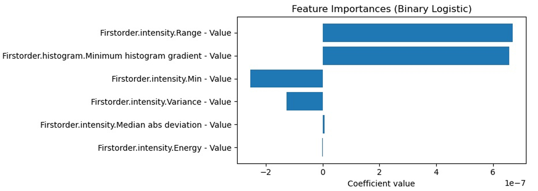
Results: CT-based sarcopenia prevalence was 45.3% using reference-based PMI thresholds. PMI declined significantly from baseline to t1 and remained stable thereafter, with women exhibiting consistently lower values. Outcome analysis showed a higher proportion of disease progression at t1 in sarcopenic patients using reference cutoffs, whereas cohort-specific quartiles demonstrated no consistent differences. Random forest models predicted sarcopenia with up to 0.73 accuracy and receiver operating characteristics area under the curve (ROC-AUC) of 0.81. LASSO regression identified the psoas short axis and cross-sectional area as the most informative features. Logistic regression using baseline radiomic features predicted disease progression status at 12 months with 0.85 accuracy, weighted F1 0.841, and AUC 0.823. Interobserver agreement for psoas measurements was high (r = 0.86).
Conclusion: Longitudinal CT-based assessment of PMI demonstrates progressive sarcopenia within the studied PDAC cohort, with sex-specific declines. Radiomic analysis of skeletal muscle provides complementary information and predictive insights, highlighting their potential to enhance the characterization of muscle status and its association with disease course in patients able to undergo repeated imaging.
Published
Issue
Section
License
Copyright (c) 2026 The authors

This work is licensed under a Creative Commons Attribution 4.0 International License.